. 2011 Jan;300(1):H262-70.
doi: 10.1152/ajpheart.00857.2010. Epub 2010 Oct 22.

Endosomal KATP channels as a reservoir after myocardial ischemia: a role for SUR2 subunits

Affiliations

Endosomal KATP channels as a reservoir after myocardial ischemia: a role for SUR2 subunits

Li Bao et al. Am J Physiol Heart Circ Physiol. 2011 Jan.

Abstract

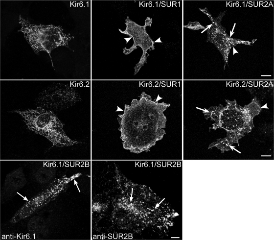
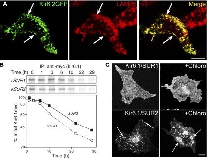
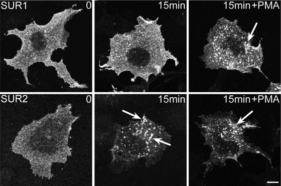
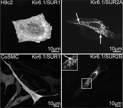
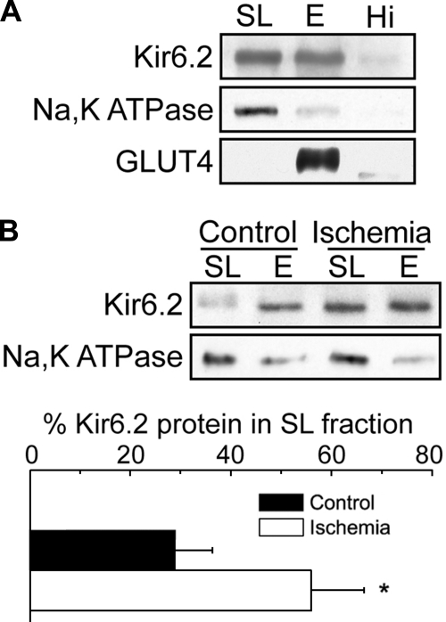
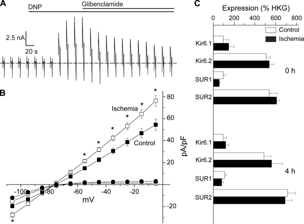
ATP-sensitive K(+) (K(ATP)) channels, composed of inward rectifier K(+) (Kir)6.x and sulfonylurea receptor (SUR)x subunits, are expressed on cellular plasma membranes. We demonstrate an essential role for SUR2 subunits in trafficking K(ATP) channels to an intracellular vesicular compartment. Transfection of Kir6.x/SUR2 subunits into a variety of cell lines (including h9c2 cardiac cells and human coronary artery smooth muscle cells) resulted in trafficking to endosomal/lysosomal compartments, as assessed by immunofluorescence microscopy. By contrast, SUR1/Kir6.x channels efficiently localized to the plasmalemma. The channel turnover rate was similar with SUR1 or SUR2, suggesting that the expression of Kir6/SUR2 proteins in lysosomes is not associated with increased degradation. Surface labeling of hemagglutinin-tagged channels demonstrated that SUR2-containing channels dynamically cycle between endosomal and plasmalemmal compartments. In addition, Kir6.2 and SUR2 subunits were found in both endosomal and sarcolemmal membrane fractions isolated from rat hearts. The balance of these K(ATP) channel subunits shifted to the sarcolemmal membrane fraction after the induction of ischemia. The K(ATP) channel current density was also increased in rat ventricular myocytes isolated from hearts rendered ischemic before cell isolation without corresponding changes in subunit mRNA expression. We conclude that an intracellular pool of SUR2-containing K(ATP) channels exists that is derived by endocytosis from the plasma membrane. In cardiac myocytes, this pool can potentially play a cardioprotective role by serving as a reservoir for modulating surface K(ATP) channel density under stress conditions, such as myocardial ischemia.


Figures

Fig. 1.
Fig. 1.
Sulfonylurea receptor (SUR) 2 targets inwardly rectifying K+ (Kir) 6.1 and Kir6.2 to intracellular vesicles. COS-1L cells were transfected with plasmids encoding Kir6.1myc (top) or Kir6.2-hemagglutinin (HA) (middle) together with SUR1 or SUR2A or in the absence of SURx (with pcDNA3). Cells were pretreated with cycloheximide for 4 h before staining for immunofluorescence microscopy (IFM). When expressed alone, Kir6.1 or Kir6.2 staining was in a reticular pattern consistent with retention in the endoplasmic reticulum. When expressed together with SUR1, both Kir6.1 and Kir6.2 were predominantly localized on plasma membranes (arrowheads). When expressed with SUR2, both Kir6.1 and Kir6.2 were detected in intracellular vesicles (arrows) and on the plasma membrane. Bottom: COS-1L cells were transfected with a 1:9 (left) or a 1:1 (right) ratio of Kir6.1:SUR2B plasmid DNA and stained for IFM using goat anti-Kir6.1 or SUR2B antibodies. The untagged Kir6.1 subunit was localized to intracellular vesicles (arrows). SUR2B was also localized to intracellular vesicles. Figure panels have been adjusted for brightness and contrast. Bars = 10 μm.
Fig. 2.
Fig. 2.
Kir6.2/SUR2A channels localize to endosomes and lysosomes, but this does not affect the protein turnover rate. A: HEK-293T cells transfected with Kir6.2-green fluorescent protein (GFP)/SUR2A cDNAs were stained for IFM with rat anti-human lysosomal-associated membrane protein-2 (LAMP2) and rhodamine-conjugated anti-rat antibody. There was extensive overlap between the GFP signal and the staining for the lysosome/late endosome marker, LAMP2 (arrows). B: HEK-293T cells transfected with plasmids encoding Kir6.1myc and either SUR1 or SUR2A were subjected to pulse-chase labeling. After immunoprecipitation (IP), samples were subjected to SDS-PAGE (B, top), and the Kir6.1myc band was quantified and plotted as a function of the chase time (B, bottom). Depicted is 1 of 3 similar experiments. C: COS-1L cells transfected with Kir6.1myc and SUR1 or SUR2A cDNAs were pretreated with chloroquine (Chloro; 100 μM) and cycloheximide for 5 h before staining for IFM with anti-myc antibodies. The drug caused the intracellular vesicles to enlarge (arrows). Figure panels have been adjusted for brightness and contrast. Bars = 10 μm.
Fig. 3.
Fig. 3.
Rapid internalization of SUR2-containing ATP-sensitive K+ (KATP) channels. COS1 cells transfected with plasmids encoding Kir6.2HA+11 and SUR1 (top) or SUR2A (bottom) were incubated for 2 h with anti-HA antibodies. Cells were warmed to 37°C for 15 min before fixation and staining with rhodamine-conjugated anti-rat antibodies. The anti-HA staining decorated the cell surface in SUR1-transfected cells, even after a 15-min incubation. By contrast, cell surface staining was initially observed in SUR2A-transfected cells, but vesicle staining was extensive after 15 min. Incubation of the cells with 100 nM PMA caused significant vesicle accumulation of antibody in SUR1-expressing cells but had little effect in SUR2-expressing cells. Figure panels have been adjusted for brightness and contrast. Bar = 10 μm.
Fig. 4.
Fig. 4.
Differential trafficking of KATP channels in cardiac myocytes and coronary artery smooth muscle cells (CoSMC). H9c2 cells and CoSMCs were transfected with the indicated plasmid combinations. IFM was performed using an anti-myc antibody. Boxed regions are depicted in the inset. Labeled vesicles (arrows) were observed in cells transfected with SUR2. Figure panels have been adjusted for brightness and contrast.
Fig. 5.
Fig. 5.
Increase in cardiac myocyte sarcolemmal KATP channels following global ischemia. A: cell fractionation was performed on rat ventricular tissue from nonischemic and ischemic rats using Optiprep gradient centrifugation. The sarcolemmal fraction (SL = 0/5% interface), endosomal fraction (E = 5/15% interface), and a loose pellet representing the highest density material (Hi) were subjected to Western blot analysis. B: representative Western blot of the SL and E fractions of membranes prepared from nonischemic control and ischemic rat hearts. The results from 3 experiments were quantified and plotted below as means ± SE. *P < 0.05. Figure panels have been adjusted for brightness and contrast.
Fig. 6.
Fig. 6.
Increase in sarcolemmal KATP channel density in ventricular myocytes isolated from ischemic rat hearts. A and B: ventricular myocytes were isolated from nonischemic or ischemic rat hearts and subjected to whole cell patch clamping with a ramp protocol. A: representative current trace is depicted. The maximal KATP channel current was estimated as the steady-state peak current after dinitrophenol (DNP) application. The dotted line indicates 0 current. B: current-voltage relationships of current densities before (circles) and after (squares) DNP application for cells in the nonischemic (filled symbols; n = 21) and ischemic (open symbols; n = 22) groups (left and right ventricular myocyte data were pooled). *P < 0.05. C: real-time RT-PCR analysis of indicted channel subunit mRNA expression in cardiac myocytes at 0 and 4 h after isolation from nonischemic (control) and ischemic hearts. Data (means ± SE) are expressed relative to housekeeping genes. Primer sequences are in the online supplement (Table S1).

Comment in

References

    1. Aguilar-Bryan L, Bryan J, Nakazaki M. Of mice and men: K(ATP) channels and insulin secretion. Recent Prog Horm Res 56:47–68, 2001 - PubMed
    1. Babenko AP, Aguilar-Bryan L, Bryan J. A view of sur/KIR6.X, KATP channels. Annu Rev Physiol 60:667–687, 1998 - PubMed
    1. Budas GR, Jovanovic S, Crawford RM, Jovanovic A. Hypoxia-induced preconditioning in adult stimulated cardiomyocytes is mediated by the opening and trafficking of sarcolemmal KATP channels. FASEB J 18:1046–1048, 2004 - PMC - PubMed
    1. Chen JW, Murphy TL, Willingham MC, Pastan I, August JT. Identification of two lysosomal membrane glycoproteins. J Cell Biol 101:85–95, 1985 - PMC - PubMed
    1. Chutkow WA, Simon MC, Le Beau MM, Burant CF. Cloning, tissue expression, and chromosomal localization of SUR2, the putative drug-binding subunit of cardiac, skeletal muscle, and vascular KATP channels. Diabetes 45:1439–1445, 1996 - PubMed

Publication types

LinkOut - more resources