

Autonomic activation links CNS oxygen toxicity to acute cardiogenic pulmonary injury
- PMID: 20971806
- PMCID: PMC3023284
- DOI: 10.1152/ajplung.00178.2010
Autonomic activation links CNS oxygen toxicity to acute cardiogenic pulmonary injury
Abstract
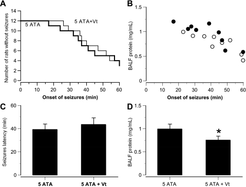
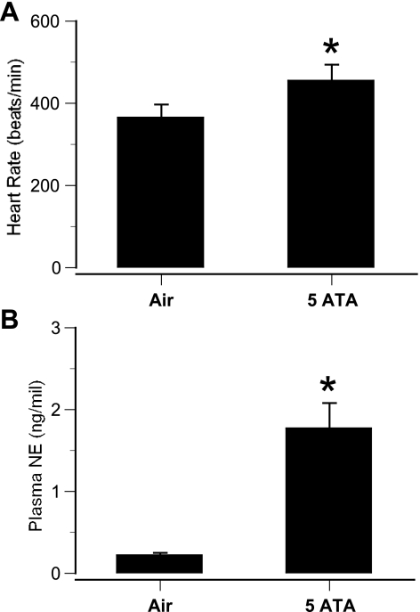
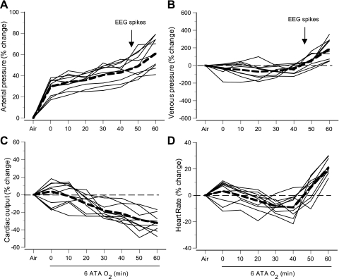
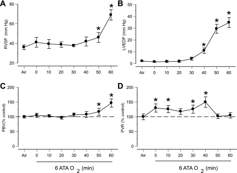
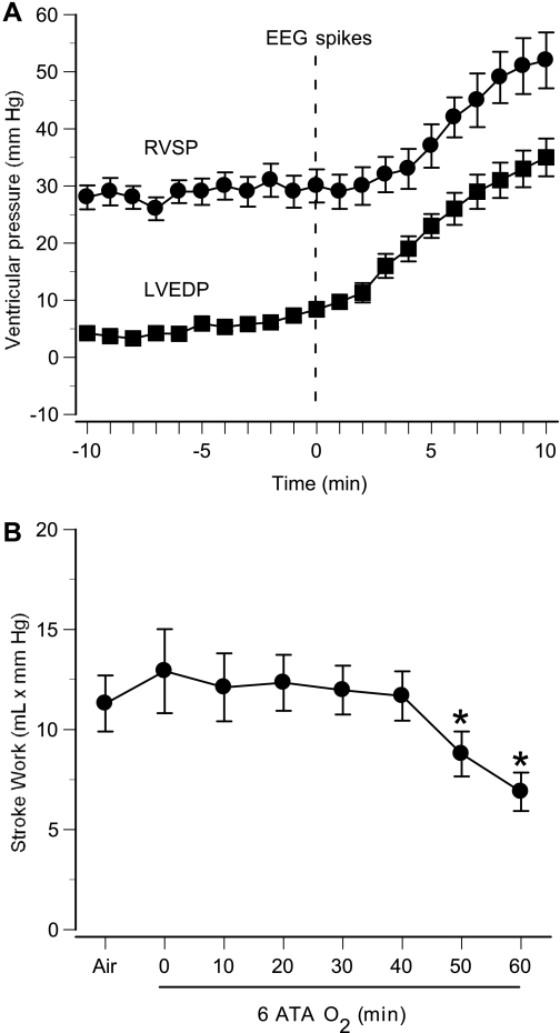
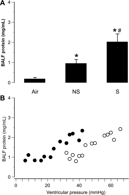
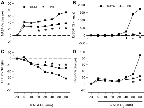
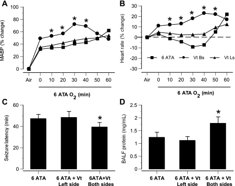
Breathing hyperbaric oxygen (HBO₂), particularly at pressures above 3 atmospheres absolute, can cause acute pulmonary injury that is more severe if signs of central nervous system toxicity occur. This is consistent with the activation of an autonomic link between the brain and the lung, leading to acute pulmonary oxygen toxicity. This pulmonary damage is characterized by leakage of fluid, protein, and red blood cells into the alveoli, compatible with hydrostatic injury due to pulmonary hypertension, left atrial hypertension, or both. Until now, however, central hemodynamic parameters and autonomic activity have not been studied concurrently in HBO₂, so any hypothetical connections between the two have remained untested. Therefore, we performed experiments using rats in which cerebral blood flow, electroencephalographic activity, cardiopulmonary hemodynamics, and autonomic traffic were measured in HBO₂ at 5 and 6 atmospheres absolute. In some animals, autonomic pathways were disrupted pharmacologically or surgically. Our findings indicate that pulmonary damage in HBO₂ is caused by an abrupt and significant increase in pulmonary vascular pressure, sufficient to produce barotrauma in capillaries. Specifically, extreme HBO₂ exposures produce massive sympathetic outflow from the central nervous system that depresses left ventricular function, resulting in acute left atrial and pulmonary hypertension. We attribute these effects on the heart and on the pulmonary vasculature to HBO₂-mediated central sympathetic excitation and catecholamine release that disturbs the normal equilibrium between excitatory and inhibitory activity in the autonomic nervous system.
Figures
Similar articles
-
Nitric oxide-mediated central sympathetic excitation promotes CNS and pulmonary O₂ toxicity.J Appl Physiol (1985). 2012 Jun;112(11):1814-23. doi: 10.1152/japplphysiol.00902.2011. Epub 2012 Mar 22. J Appl Physiol (1985). 2012. PMID: 22442027 Free PMC article.
-
Baroreceptor afferents modulate brain excitation and influence susceptibility to toxic effects of hyperbaric oxygen.J Appl Physiol (1985). 2014 Sep 1;117(5):525-34. doi: 10.1152/japplphysiol.00435.2014. Epub 2014 Jul 3. J Appl Physiol (1985). 2014. PMID: 24994889
-
Complications and side effects of hyperbaric oxygen therapy.Aviat Space Environ Med. 2000 Feb;71(2):119-24. Aviat Space Environ Med. 2000. PMID: 10685584
-
Effect of hyperbaric oxygenation on brain hemodynamics, hemoglobin oxygenation and mitochondrial NADH.Brain Res Rev. 2007 Jun;54(2):294-304. doi: 10.1016/j.brainresrev.2007.04.004. Epub 2007 Apr 27. Brain Res Rev. 2007. PMID: 17570266 Review.
-
Hyperbaric oxygen in the treatment of acute ischemic stroke: an unsettled issue.J Neurol Sci. 1997 Sep 1;150(1):27-31. doi: 10.1016/s0022-510x(97)05398-7. J Neurol Sci. 1997. PMID: 9260854 Review.
Cited by
-
Nitric oxide-mediated central sympathetic excitation promotes CNS and pulmonary O₂ toxicity.J Appl Physiol (1985). 2012 Jun;112(11):1814-23. doi: 10.1152/japplphysiol.00902.2011. Epub 2012 Mar 22. J Appl Physiol (1985). 2012. PMID: 22442027 Free PMC article.
-
The O2-sensitive brain stem, hyperoxic hyperventilation, and CNS oxygen toxicity.Front Physiol. 2022 Jul 26;13:921470. doi: 10.3389/fphys.2022.921470. eCollection 2022. Front Physiol. 2022. PMID: 35957982 Free PMC article. Review.
-
Exogenous ketone salts inhibit superoxide production in the rat caudal solitary complex during exposure to normobaric and hyperbaric hyperoxia.J Appl Physiol (1985). 2021 Jun 1;130(6):1936-1954. doi: 10.1152/japplphysiol.01071.2020. Epub 2021 Mar 4. J Appl Physiol (1985). 2021. PMID: 33661724 Free PMC article.
-
Plasma Proteomics-Based Discovery of Mechanistic Biomarkers of Hyperbaric Stress and Pulmonary Oxygen Toxicity.Metabolites. 2023 Aug 23;13(9):970. doi: 10.3390/metabo13090970. Metabolites. 2023. PMID: 37755249 Free PMC article.
-
Does Heart Rate Variability Predict Impairment of Operational Performance in Divers?Sensors (Basel). 2024 Dec 3;24(23):7726. doi: 10.3390/s24237726. Sensors (Basel). 2024. PMID: 39686263 Free PMC article.
References
-
- Abel FL, McNamee JE, Cone DL, Clarke D, Tao J. Effects of hyperbaric oxygen on ventricular performance, pulmonary blood volume, and systemic and pulmonary vascular resistance. Undersea Hyperb Med 27: 67–73, 2000 - PubMed
-
- Abel FL, Waldhausen JA, Daly WJ, Pearce WL. Pulmonary blood volume in hemorrhagic shock in the dog and primate. Am J Physiol 213: 1072–1078, 1967 - PubMed
-
- Balentine JD. Pathology of Oxygen Toxicity. New York: Academic, 1982
-
- Baumann A, Audibert G, McDonnell J, Mertes PM. Neurogenic pulmonary edema. Acta Anaesthesiol Scand 51: 447–455, 2007 - PubMed
-
- Bean J, Zee D, Thom B. Pulmonary changes with convulsions induced by drugs and oxygen at high pressure. J Appl Physiol 21: 865–872, 1966 - PubMed
Publication types
MeSH terms
Substances
Grants and funding
LinkOut - more resources
Full Text Sources