. 2010 Dec;16(12):2455-63.
doi: 10.1261/rna.2334110. Epub 2010 Oct 22.

Deciphering the role of glucosamine-6-phosphate in the riboswitch action of glmS ribozyme

Affiliations

Deciphering the role of glucosamine-6-phosphate in the riboswitch action of glmS ribozyme

Yao Xin et al. RNA. 2010 Dec.

Abstract

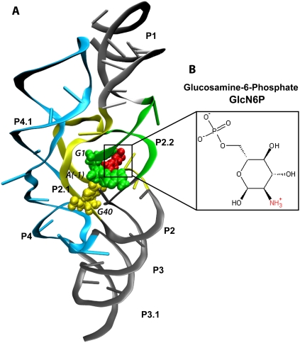
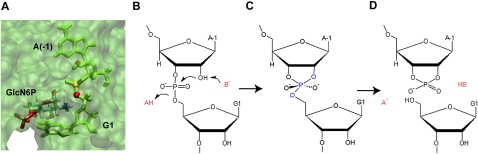
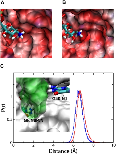
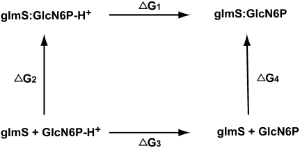
The GlmS ribozyme is believed to exploit a general acid-base catalytic mechanism in the presence of glucosamine-6-phosphate (GlcN6P) to accelerate self-cleavage by approximately six orders of magnitude. The general acid and general base are not known, and the role of the GlcN6P cofactor is even less well understood. The amine group of GlcN6P has the ability to either accept or donate a proton and could therefore potentially act as an acid or a base. In order to decipher the role of GlcN6P in the self-cleavage of glmS, we have determined the preferred protonation state of the amine group in the wild-type and an inactive G40A mutant using molecular dynamics simulations and free energy calculations. Here we show that, upon binding of GlcN6P to wild-type glmS, the pK(a) of the amine moiety is altered by the active site environment, decreasing by about 2.2 from a solution pK(a) of about 8.2. On the other hand, we show that the pK(a) of the amine group slightly increases to about 8.4 upon binding to the G40A inactive mutant of glmS. These results suggest that GlcN6P acts as a general acid in the self-cleavage of glmS. Upon binding to glmS, GlcN6P can easily release a proton to the 5'-oxygen of G1 during self-cleavage of the backbone phosphodiester bond. However, in the G40A inactive mutant of glmS, the results suggest that the ability of GlcN6P to easily release its proton is diminished, in addition to the possible lack of G40 as an effective base.


Figures

FIGURE 1.
FIGURE 1.
X-ray crystal structure of glmS ribozyme (A) from Thermoanaerobacter tengcongensis complexed with GlcN6P (B), shown in red. Loop and helix segments are labeled according to Klein et al. (2007b; Fig. 1C). glmS self-cleaves its backbone phosphodiester bond between A(-1) and G1, shown in green. G40 is shown in yellow.
FIGURE 2.
FIGURE 2.
The active site and the proposed mechanism for the self-cleavage of the glmS ribozyme. A view of the active site (A) of the glmS ribozyme with A(-1) and G1 shown in green, 2′ O of A(-1) shown as red sphere, the scissile P shown as yellow, and 5′ O of G1 also shown as red sphere. The precleavage (B), transition (C), and post-cleavage (D) states of the cleavage site with the general acid, HA, and base, B are also shown.
FIGURE 3.
FIGURE 3.
Normalized probability distribution, p(r), of important atomic distances and angle in the binding site of both the wild-type (blue lines) and G40A inactive mutant (red lines) of the glmS ribozyme. (A) Distance from GlcN6P:N to G1:O5′. (B) Distance from A(-1):O2′ to scissile P (P*). (C) Angle between A(-1):O2′, P*, and G1:O5′. (D) Distance from G40:N1 to A(-1):O2′ (A40:N1 to A[-1]:O2′ in the G40A inactive mutant). The insets in A to D are the atomic view of these distances and angles. The cleavage site A(-1) and G1, the metabolite GlcN6P, and residue 40 are shown and colored by element. The rest of the active site is shown as white surface. The functional essential atoms are labeled (green) within the figure.
FIGURE 4.
FIGURE 4.
Hydration density of the binding site of the wild type (A) and G40A inactive mutant (B) of the glmS ribozyme. GlcN6P is shown using ball and stick and colored by element. WAT1 and WAT2 are the two highly localized and conserved water molecules in the binding site. Red-colored mesh represents 10 times that of bulk water. Green-colored mesh shows six times that of bulk water. Shown using a green sphere is a Mg2+ ion in the binding site that helps to stabilize the complex.
FIGURE 5.
FIGURE 5.
Electrostatic potential map of the binding site of the wild-type (A) and G40A inactive mutant (B) of the glmS ribozyme. The normalized probability distribution, p(r), of the distance between GlcN6P:N and G40:N1 of the wild-type glmS (blue line) and the distance between GlcN6P:N and A40:N1 of the G40A inactive mutant (red line). (C) The electrostatic maps are colored by potential with a range from −10 kT/e to +10 kT/e. Red color denotes negative potential and blue color denotes positive potential.
FIGURE 6.
FIGURE 6.
Thermodynamic cycle connecting the deprotonation of GlcN6P in water and in the binding site of the glmS ribozyme.

Similar articles

Cited by

References

    1. Allison S, Xin Y 2006. Electrokinetic transport of a spherical gel-layer model particle: Inclusion of charge regulation and application to polystyrene sulfonate. J Colloid Interface Sci 299: 977–988 - PubMed
    1. Anderson CF, Record MT 1995. Salt nucleic-acid interactions. Annu Rev Phys Chem 46: 657–700 - PubMed
    1. Baker NA, Sept D, Joseph S, Holst MJ, McCammon JA 2001. Electrostatics of nanosystems: Application to microtubules and the ribosome. Proc Natl Acad Sci 98: 10037–10041 - PMC - PubMed
    1. Banas P, Walter NG, Sponer J, Otyepka M 2010. Protonation states of the key active site residues and structural dynamics of glmS riboswitch as reveled by molecular dynamics. J Phys Chem B 114: 8701–8712 - PMC - PubMed
    1. Barrick JE, Corbino KA, Winkler WC, Nahvi A, Mandal M, Collins J, Lee M, Roth A, Sudarsan N, Jona I, et al. 2004. New RNA motifs suggest an expanded scope for riboswitches in bacterial genetic control. Proc Natl Acad Sci 101: 6421–6426 - PMC - PubMed

Publication types

MeSH terms

LinkOut - more resources