. 2010 Nov 15;24(22):2505-16.
doi: 10.1101/gad.605910. Epub 2010 Oct 22.

Rec8-containing cohesin maintains bivalents without turnover during the growing phase of mouse oocytes

Affiliations

Rec8-containing cohesin maintains bivalents without turnover during the growing phase of mouse oocytes

Kikuë Tachibana-Konwalski et al. Genes Dev. .

Abstract

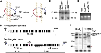
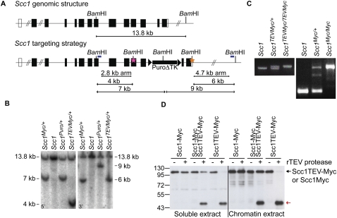
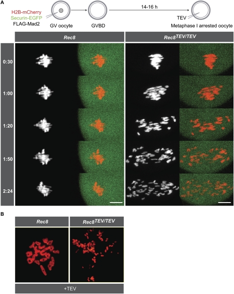
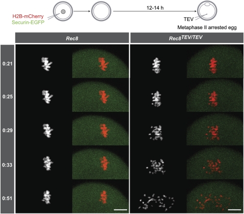
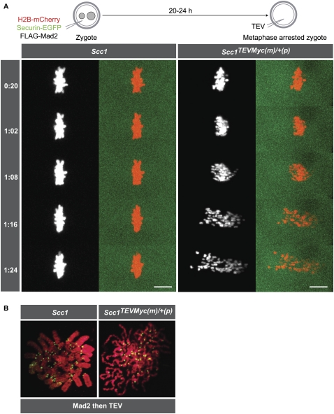
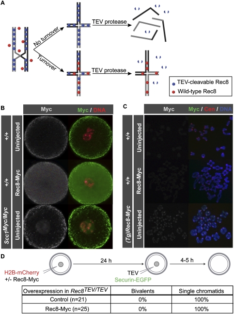
During female meiosis, bivalent chromosomes are thought to be held together from birth until ovulation by sister chromatid cohesion mediated by cohesin complexes whose ring structure depends on kleisin subunits, either Rec8 or Scc1. Because cohesion is established at DNA replication in the embryo, its maintenance for such a long time may require cohesin turnover. To address whether Rec8- or Scc1-containing cohesin holds bivalents together and whether it turns over, we created mice whose kleisin subunits can be cleaved by TEV protease. We show by microinjection experiments and confocal live-cell imaging that Rec8 cleavage triggers chiasmata resolution during meiosis I and sister centromere disjunction during meiosis II, while Scc1 cleavage triggers sister chromatid disjunction in the first embryonic mitosis, demonstrating a dramatic transition from Rec8- to Scc1-containing cohesin at fertilization. Crucially, activation of an ectopic Rec8 transgene during the growing phase of Rec8(TEV)(/TEV) oocytes does not prevent TEV-mediated bivalent destruction, implying little or no cohesin turnover for ≥2 wk during oocyte growth. We suggest that the inability of oocytes to regenerate cohesion may contribute to age-related meiosis I errors.


Figures

Figure 1.
Figure 1.
Generation of a TEV-cleavable Rec8 allele. (A) Schematic of the cohesin ring consisting of an Smc1/Smc3 heterodimer bridged by the α-kleisin subunit. TEV protease-mediated cleavage of an α-kleisin subunit engineered to contain TEV protease recognition sites allows inducible opening of the ring. (B) Targeting strategy for generating a Rec8TEV allele. Schematic of the Rec8 genomic locus and targeted allele (Rec8TEV). Translated exons are shown as filled boxes. The pink star indicates three tandem TEV protease recognition sites, including a novel MfeI site, that were inserted into exon 10. The puromycin-truncated thymidine kinase selection cassette (rectangle) is flanked by LoxP sites (triangles). The relative location of the 5′ and 3′ probes (blue bars) and MfeI sites that were used in Southern blot analysis are shown on the targeted locus. (C) Southern blot analysis of wild-type (Rec8) and heterozygous (Rec8TEV/+) mouse ES genomic DNA (gDNA) digested with MfeI and hybridized with 5′ and 3′ probes to check for homologous recombination. (D) PCR analysis of wild-type (Rec8), heterozygous (Rec8TEV/+), and homozygous (Rec8TEV/TEV) mouse ear gDNA. (E) Western blot analysis of Rec8 cleaved by recombinant TEV protease (rTEV) in testes chromatin extracts from wild-type and Rec8TEV/TEV mice. Rec8 was detected by anti-Rec8 antibody. The red arrow indicates the Rec8 cleavage fragment.
Figure 2.
Figure 2.
Generation of a TEV-cleavable Scc1 allele. (A) Targeting strategy for generating an Scc1TEVMyc allele. Schematic of the Scc1 genomic locus and targeted alleles (Scc1TEVMyc or Scc1Myc). The pink star indicates three tandem TEV protease recognition sites, including a novel BamHI restriction site, that were inserted into exon 11. The orange star indicates a c-Myc10 epitope, including a novel BamHI restriction site that was inserted before the Stop codon. The relative location of the 5′ and 3′ probes (blue bars) and BamHI sites that were used in Southern blot analysis are shown on the targeted locus. (B) Southern blot analysis of wild-type (Scc1) and heterozygous (Scc1TEVMyc/+ and Scc1Myc/+) mouse ES gDNA digested with BamHI and hybridized with 5′ and 3′ probes to check for homologous recombination. (C) PCR analysis of wild-type (Scc1), heterozygous (Scc1TEVMyc/+), and homozygous (Scc1TEVMyc/TEVMyc) mouse ear gDNA (left panel), and wild-type (Scc1), heterozygous (Scc1Myc/+), and homozygous (Scc1Myc/Myc) mouse ear gDNA (right panel). (D) Western blot analysis of Scc1 cleavage by recombinant TEV protease in soluble and chromatin extracts from Scc1Myc/Myc, Scc1Myc/TEVMyc, and Scc1TEVMyc/TEVMyc ES cells. Scc1TEV-Myc and Scc1-Myc were detected by anti-c-Myc antibody. The red arrow indicates the Scc1 cleavage fragment.
Figure 3.
Figure 3.
Rec8 cleavage triggers chiasmata resolution and sister centromere disjunction of bivalent chromosomes. (A) Wild-type and Rec8TEV/TEV oocytes expressing H2B-mCherry, Securin-EGFP, and Flag-Mad2 were cultured for 14–16 h. Metaphase I-arrested oocytes were injected with TEV protease mRNA (time 0), and chromosome movements were visualized by time-lapse confocal microscopy (h:mm). Still images from a representative movie of each genotype are shown. The left panels show the H2B-mCherry channel and the right panels show H2B-mCherry and Securin-EGFP pseudocolored in red and green, respectively. Bar, 10 μm. (B) Wild-type and Rec8TEV/TEV oocytes expressing TEV protease were matured in vitro for 5 h. Chromosome spreads were prepared and stained with Hoechst to visualize DNA (red) and CREST to mark centromeres (green).
Figure 4.
Figure 4.
Rec8 cleavage triggers sister centromere disjunction in meiosis II. Wild-type and Rec8TEV/TEV oocytes expressing H2B-mCherry and Securin-EGFP were cultured for 12–14 h. Metaphase II-arrested eggs were injected with TEV protease (time 0), and chromosome movements were visualized by time-lapse confocal microscopy (h:mm). Bar, 10 μm.
Figure 5.
Figure 5.
Scc1 cleavage triggers sister chromatid disjunction in the first embryonic mitosis. (A) Wild-type and Scc1TEVMyc/TEVMyc females were mated to wild-type males, and wild-type and Scc1TEVMyc(m)/+(p) zygotes (maternal vs. paternal alleles) were isolated, respectively. Zygotes expressing H2B-mCherry, Securin-EGFP, and Flag-Mad2 were cultured for 20–24 h. Metaphase-arrested zygotes were injected with TEV protease mRNA (time 0), and chromosome movements were visualized by time-lapse confocal microscopy (h:mm). Bar, 10 μm. (B) Wild-type and Scc1TEVMyc(m)/+(p) zygotes expressing Flag-Mad2 were cultured for 20–24 h. Metaphase-arrested zygotes were injected with TEV protease mRNA and cultured for at least 2 h. Chromosome spreads were prepared and stained with DAPI to visualize DNA (red) and CREST to mark centromeres (green).
Figure 6.
Figure 6.
No cohesin turnover in mature GV oocytes. (A) Schematic of cohesin turnover assay. TEV-cleavable Rec8 (blue spheres) is loaded during meiotic DNA replication and maintains the bivalent structure. If cohesin does not turn over following cohesion establishment and recombination, then wild-type Rec8 (red spheres) expressed post-recombination fails to be incorporated into functional cohesin complexes and TEV cleavage resolves bivalent chromosomes into single chromatids. If, on the other hand, cohesin turns over, then wild-type Rec8 expressed post-recombination forms de novo cohesion and the bivalent structure becomes resistant to TEV cleavage. (B) Wild-type GV oocytes were either injected with Rec8-Myc mRNA (middle panel) or uninjected (top panel) and cultured in the presence of IBMX for 24 h prior to fixation. (Bottom panel) An Scc1Myc/Myc GV oocyte was included as an uninjected control for c-Myc staining. Oocyte in situ immunostaining was performed using c-Myc antibody to visualize either overexpressed Rec8-Myc or endogenous Scc1-Myc (green), and DNA was stained with Hoechst (red). (C) Wild-type GV oocytes were either injected with Rec8-Myc mRNA (middle panel) or uninjected (top panel) and cultured in the presence of IBMX for 24 h. Oocytes were then cultured in IBMX-free medium for 5 h. Chromosome spreads were prepared and stained with c-Myc antibody to visualize either overexpressed Rec8-Myc or Rec8-Myc expressed from a constitutive BAC transgene, (Tg)Rec8-Myc (green), CREST to mark centromeres (red), and Hoechst to visualize DNA (red). (D) Rec8TEV/TEV oocytes expressing H2B-mCherry with or without Rec8-Myc were cultured in the presence of IBMX for 24 h and then injected with TEV protease and Securin-EGFP mRNA. Oocytes were released into IBMX-free culture medium and chromosomes were visualized by time-lapse confocal microscopy. Oocytes were scored as containing either 20 bivalent chromosomes or at least 72 single chromatids (and no bivalents) by 5 h post-GVBD.
Figure 7.
Figure 7.
No cohesin turnover in growing-phase oocytes. (A) Western blot analysis of Rec8-Myc using c-Myc antibody on whole ovary and testis extracts. Rec8-Myc is expressed constitutively from either a multicopy (Tg)Rec8-Myc BAC transgene (second row) or a (Tg)Stop/Rec8-Myc BAC transgene carrying a conditional Stop cassette flanked by LoxP sites (third row) that is activated by Cre recombinase under the control of the Sox2 (fourth row) or Zp3 (fifth row) promoter. The asterisks indicate cross-reacting bands with c-Myc antibody. (B) Chromosome spreads were prepared from four types of oocytes at 5 h post-GVBD. Chromosome spreads were stained with c-Myc antibody to visualize Rec8-Myc (green), CREST to mark centromeres (red), and Hoechst to visualize DNA (blue). (C) Seven types of GV oocytes expressing TEV protease, H2B-mCherry, and Securin-EGFP were cultured in the presence of IBMX for 1 h, then released into IBMX-free culture medium, and chromosomes were visualized by time-lapse confocal microscopy. Oocytes (n = number of cells) were scored as containing either 20 bivalent chromosomes or at least 72 single chromatids (and no bivalents) by 5 h post-GVBD.

Comment in

References

    1. Adams DJ, Quail MA, Cox T, van der Weyden L, Gorick BD, Su Q, Chan WI, Davies R, Bonfield JK, Law F, et al. 2005. A genome-wide, end-sequenced 129Sv BAC library resource for targeting vector construction. Genomics 86: 753–758 - PubMed
    1. Bannister LA, Reinhold LG, Munroe RJ, Schimenti JC 2004. Positional cloning and characterization of mouse mei8, a disrupted allele of the meiotic cohesin Rec8. Genesis 40: 184–194 - PubMed
    1. Buonomo SB, Clyne RK, Fuchs J, Loidl J, Uhlmann F, Nasmyth K 2000. Disjunction of homologous chromosomes in meiosis I depends on proteolytic cleavage of the meiotic cohesin Rec8 by separin. Cell 103: 387–398 - PubMed
    1. Días-Martínez LA, Giménez-Abián JF, Clarke DJ 2007. Cohesin is dispensable for centromere cohesion in human cells. PLoS ONE 2: e318 doi: 10.1371/journal.pone.0000318 - PMC - PubMed
    1. Días-Martínez LA, Giménez-Abián JF, Clarke DJ 2008. Chromosome cohesion—Rings, knots, orcs and fellowship. J Cell Sci 121: 2107–2114 - PubMed

Publication types