. 2010 Dec 24;285(52):40785-92.
doi: 10.1074/jbc.M110.173161. Epub 2010 Oct 22.

A single amino acid in human APOBEC3F alters susceptibility to HIV-1 Vif

Affiliations

A single amino acid in human APOBEC3F alters susceptibility to HIV-1 Vif

John S Albin et al. J Biol Chem. .

Abstract

Human APOBEC3F (huA3F) potently restricts the infectivity of HIV-1 in the absence of the viral accessory protein virion infectivity factor (Vif). Vif functions to preserve viral infectivity by triggering the degradation of huA3F but not rhesus macaque A3F (rhA3F). Here, we use a combination of deletions, chimeras, and systematic mutagenesis between huA3F and rhA3F to identify Glu(324) as a critical determinant of huA3F susceptibility to HIV-1 Vif-mediated degradation. A structural model of the C-terminal deaminase domain of huA3F indicates that Glu(324) is a surface residue within the α4 helix adjacent to residues corresponding to other known Vif susceptibility determinants in APOBEC3G and APOBEC3H. This structural clustering suggests that Vif may bind a conserved surface present in multiple APOBEC3 proteins.


Figures
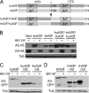
FIGURE 1.
FIGURE 1.
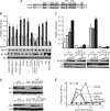
Susceptibility of huA3F to HIV-1 Vif maps to the huA3F CTD. A, schematic depiction of the chimeras used in B. B, cotransfection experiment demonstrating the instability in the presence of HIV-1 Vif of chimeras between rhA3F and huA3F that contain the huA3F CTD. C and D, cotransfection experiments demonstrating that the CTDs of huA3F and rhA3F are destabilized by HIV-1 Vif or SIV Vif, respectively, whereas the corresponding NTDs remains highly expressed.
FIGURE 2.
FIGURE 2.
Substitution of rhA3F residues at positions 323–324 of huA3F results in phenotypic Vif resistance. A, schematic depiction of the CTDs of huA3F and rhA3F with relevant amino acids shown. B, single-cycle infectivity data quantifying the effects of HIV-1 Vif on the rescue of HIV-1 infectivity. All mutants remained competent for restriction. Relative infectivity represents the mean ± S.E. of four independent experiments. Western blots demonstrating intracellular APOBEC3 and Vif levels corresponding to one of these experiments are shown below. C, single-cycle infectivity data demonstrating the continued restriction of HIV-1 by untagged huA3F Q323E/E324K and huA3G D128K/D130K in the presence of increasing amounts of Vif. Relative infectivity represents the mean ± S.E. (error bars) of two independent experiments done in duplicate. The Western blots demonstrating the stability of Vif-resistant huA3F and huA3G variants in the presence of Vif correspond to one of these infectivity experiments. D, Western blots showing the expression levels of huA3F, huA3G, and their Vif-resistant variants in the SupT11 cell lines used in E. E, representative spreading infection curves demonstrating that wild-type HIV-1IIIB is restricted by Vif-resistant variants huA3F Q323E/E324K and huA3G D128K/D130K but not the corresponding wild-type APOBEC3 proteins. Open symbols indicate vector control or huA3F- or huA3G-expressing cell lines. Filled symbols indicate cell lines expressing huA3F Q323E/E324K or huA3G D128K/D130K. The x axis is offset from zero to permit visualization of curves yielding no detectable spread.
FIGURE 3.
FIGURE 3.
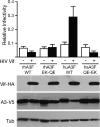
Substitution of human residues at positions 323–324 of rhA3F does not sensitize rhA3F to HIV-1 Vif. Single-cycle infectivity experiments demonstrate that substituting the human residues at positions 323–324 of rhA3F does not sensitize this restriction factor to permit infectivity recovery in the presence of Vif. Data represent the mean and S.E. (error bars) of three independent experiments. Western blots corresponding to one of the single-cycle experiments shown demonstrate the correlation between intracellular stability of APOBEC3 variants and functional recovery in infectivity.
FIGURE 4.
FIGURE 4.
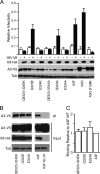
The identity of residue 324 is a primary determinant of the degradation sensitivity of huA3F to HIV-1 Vif. A, single-cycle infectivity data quantifying the restriction and Vif-sensitivity phenotypes of single and double human-to-rhesus and human-to-alanine mutations at positions 323 and/or 324 of huA3F, where only mutations at position 324 ablate Vif responsiveness. Data represent the mean ± S.E. (error bars) of three independent experiments. Western blots corresponding to one of these experiments demonstrating that mutations at position 324 of huA3F result in resistance to Vif-mediated degradation are shown below. B, representative experiment demonstrating the lack of effect of mutations at huA3F positions 323–324 on coimmunoprecipitation with HA-tagged Vif. The bands in each row are taken unaltered from different parts of the same blot. C, quantification of the results from a total of five independent experiments including the one shown in B. Relative binding represents the ratio of IP V5 signal to cellular V5 signal normalized to the ratio observed for A3F in a given experiment (set to 1).
FIGURE 5.
FIGURE 5.
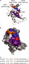
Model structure of the CTD of huA3F. A, ribbon diagram depicting the CTD of huA3F. The region encompassing the huA3F equivalents of all known single amino acid determinants of Vif sensitivity is shown in blue, with Asp311, Asp313, and Glu316 shown in orange and Glu324 in red. The region previously implicated in huA3F interaction with Vif (residues 283–300) is colored purple. B, predicted surface of the huA3F CTD. C, alignment of residues in the α4 helix encompassing known determinants of Vif susceptibility.

References

    1. Romani B., Engelbrecht S., Glashoff R. H. (2009) Arch. Virol. 154, 1579–1588 - PubMed
    1. Henriet S., Mercenne G., Bernacchi S., Paillart J. C., Marquet R. (2009) Microbiol. Mol. Biol. Rev. 73, 211–232 - PMC - PubMed
    1. Albin J. S., Harris R. S. (2010) Expert Rev. Mol. Med. 12, 1–26 - PMC - PubMed
    1. Mangeat B., Turelli P., Liao S., Trono D. (2004) J. Biol. Chem. 279, 14481–14483 - PubMed
    1. Bogerd H. P., Doehle B. P., Wiegand H. L., Cullen B. R. (2004) Proc. Natl. Acad. Sci. U.S.A. 101, 3770–3774 - PMC - PubMed

Publication types

MeSH terms

Substances