. 2010 Dec 24;285(52):40573-80.
doi: 10.1074/jbc.M110.173658. Epub 2010 Oct 22.

The rate of folding dictates substrate secretion by the Escherichia coli hemolysin type 1 secretion system

Affiliations

The rate of folding dictates substrate secretion by the Escherichia coli hemolysin type 1 secretion system

Patrick J Bakkes et al. J Biol Chem. .

Abstract

Secretion of the Escherichia coli toxin hemolysin A (HlyA) is catalyzed by the membrane protein complex HlyB-HlyD-TolC and requires a secretion sequence located within the last 60 amino acids of HlyA. The Hly translocator complex exports a variety of passenger proteins when fused N-terminal to this secretion sequence. However, not all fusions are secreted efficiently. Here, we demonstrate that the maltose binding protein (MalE) lacking its natural export signal and fused to the HlyA secretion signal is poorly secreted by the Hly system. We anticipated that folding kinetics might be limiting secretion, and we therefore introduced the "folding" mutation Y283D. Indeed this mutant fusion protein was secreted at a much higher level. This level was further enhanced by the introduction of a second MalE folding mutation (V8G or A276G). Secretion did not require the molecular chaperone SecB. Folding analysis revealed that all mutations reduced the refolding rate of the substrate, whereas the unfolding rate was unaffected. Thus, the efficiency of secretion by the Hly system is dictated by the folding rate of the substrate. Moreover, we demonstrate that fusion proteins defective in export can be engineered for secretion while still retaining function.


Figures

FIGURE 1.
FIGURE 1.
Schematic view of the toxin HlyA and the proteins used in this study. A, shown is HlyA with its C-terminal secretion signal (blue). N and C represent the N and C terminus of the protein, respectively. The putative hydrophobic membrane insertion domain (M) and the Ca2+-binding motifs are shown in red and green, respectively. For toxin activity, HlyA requires acylation of the two indicated lysine residues. B, a representation of the HlyAc fusion proteins is shown. A fragment consisting of the C-terminal amino acids 805–1023 of HlyA (HlyAc) is fused to a protein of interest via a flexible linker sequence (purple). A tobacco etch virus protease (TEV) and factor Xa cleavage site permits the specific removal of the N-terminal His6-tag and the HlyAc fragment, respectively. For more details, see the supplemental data. C, schematic view of the HlyAc fragment (left panel) that is secreted at high level as determined by SDS-PAGE (right panel). Analysis of the culture supernatant (no TCA precipitation was performed) of E. coli cells harboring pK184-HlyBD and pHlyAc. Cells were grown in ZYM-5052 medium (34) containing lactose that allows autoinduction of expression of hlyB and hlyD. At an A600 of 0.8, the expression of hlyAc was induced by the addition of arabinose to a final concentration of 10 mm. Protein was stained with CBB. M, marker proteins with indicated molecular masses (kDa).
FIGURE 2.
FIGURE 2.
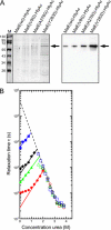
Secretion and folding of the MalE(WT)-HlyAc fusion protein and its variants. A, cells containing pK184-HlyBD and a plasmid encoding MalE-HlyAc fusion protein as indicated were grown in LB medium supplemented with 1.5 mm isopropyl 1-thio-β-d-galactopyranoside to induce hlyBD coexpression. At A660 ∼ 0.8, the production of the MalE-HlyAc fusion proteins was induced by the addition of arabinose to a final concentration of 10 mm arabinose. Five hours after induction, protein in the culture supernatants was TCA-precipitated and analyzed by SDS-PAGE with CBB staining (left panel) or immunoblotting using HlyA-specific polyclonal antibodies (right panel). B, shown is a chevron plot displaying relaxation times for the refolding and unfolding of the MalE-HlyAc fusion proteins at different urea concentrations. Relaxation times for refolding (closed symbols) and unfolding (open symbols) of wild-type (circles), V8G (triangles), A276G (diamonds), and Y283D (squares) fusion protein are indicated. The intersection of the dashed and solid lines with the y axis indicate the relaxation time for unfolding and refolding at zero denaturant concentration, respectively (see also Table 1).
FIGURE 3.
FIGURE 3.
Secretion and functional activity of the MalE(V8G/Y283D)-HlyAc fusion protein. A, production of HlyAc and the various fusion proteins was performed as described in the legend to Fig. 2. Five hours after induction, protein in the culture supernatants was TCA-precipitated and analyzed by SDS-PAGE (left panel) or immunoblotting using HlyA-specific polyclonal antibodies (right panel). M, marker proteins with indicated molecular weights (kDa). B, secreted MalE(V8G/Y283D)-HlyAc was purified by immobilized metal ion affinity chromatography, treated with factor Xa where indicated, and then applied to an amylose resin. Nonspecifically bound protein was removed by extensive washing, and the bound protein was eluted with buffer containing 10 mm maltose and analyzed by SDS-PAGE, followed by CBB staining. I, input; FT, flow through; E, elution with maltose; M, marker proteins with indicated molecular masses (kDa). A, the arrow indicates the position of the MaIE-fusion protein. B, the upper arrow indicates the MaIE-fusion protein, whereas the lower arrow indicates the MaIE core protein.
FIGURE 4.
FIGURE 4.
Secretion of the MalE-HlyAc fusion proteins by cells lacking SecB. E. coli MC4100 ΔsecB::CmR cells containing pK184-HlyBD and a plasmid encoding the MalE-HlyAc fusion protein or the HlyAc fragment were grown in LB medium supplemented with 1.5 mm isopropyl 1-thio-β-d-galactopyranoside for hlyBD coexpression. At A660 ∼ 0.8, the production of HlyAc and the different MalE-HlyAc fusion proteins as indicated was induced by the addition of 10 mm arabinose. Before (−) and after induction, at the time points indicated, protein in the culture supernatants was TCA-precipitated and analyzed by immunoblotting using HlyA-specific polyclonal antibodies.

References

    1. Holland I. B., Schmitt L., Young J. (2005) Mol. Membr. Biol. 22, 29–39 - PubMed
    1. Wickner W., Schekman R. (2005) Science 310, 1452–1456 - PubMed
    1. Davidson A. L., Chen J. (2004) Annu. Rev. Biochem. 73, 241–268 - PubMed
    1. Koronakis V., Sharff A., Koronakis E., Luisi B., Hughes C. (2000) Nature 405, 914–919 - PubMed
    1. Johnson J. M., Church G. M. (1999) J. Mol. Biol. 287, 695–715 - PubMed

Publication types

MeSH terms

LinkOut - more resources