Structural insight into the differential effects of omega-3 and omega-6 fatty acids on the production of Abeta peptides and amyloid plaques
- PMID: 20971855
- PMCID: PMC3057860
- DOI: 10.1074/jbc.M110.183608
Structural insight into the differential effects of omega-3 and omega-6 fatty acids on the production of Abeta peptides and amyloid plaques
Abstract
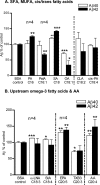
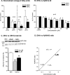
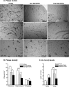
Several studies have shown the protective effects of dietary enrichment of various lipids in several late-onset animal models of Alzheimer Disease (AD); however, none of the studies has determined which structure within a lipid determines its detrimental or beneficial effects on AD. High-sensitivity enzyme-linked immunosorbent assay (ELISA) shows that saturated fatty acids (SFAs), upstream omega-3 FAs, and arachidonic acid (AA) resulted in significantly higher secretion of both Aβ 40 and 42 peptides compared with long chain downstream omega-3 and monounsaturated FAs (MUFA). Their distinct detrimental action is believed to be due to a structural template found in their fatty acyl chains that lack SFAs, upstream omega-3 FAs, and AA. Immunoblotting experiments and use of APP-C99-transfected COS-7 cells suggest that FA-driven altered production of Aβ is mediated through γ-secretase cleavage of APP. An early-onset AD transgenic mouse model expressing the double-mutant form of human amyloid precursor protein (APP); Swedish (K670N/M671L) and Indiana (V717F), corroborated in vitro findings by showing lower levels of Aβ and amyloid plaques in the brain, when they were fed a low fat diet enriched in DHA. Our work contributes to the clarification of aspects of structure-activity relationships.
Figures


Similar articles
-
Detrimental effects of arachidonic acid and its metabolites in cellular and mouse models of Alzheimer's disease: structural insight.Neurobiol Aging. 2012 Apr;33(4):831.e21-31. doi: 10.1016/j.neurobiolaging.2011.07.014. Epub 2011 Sep 13. Neurobiol Aging. 2012. PMID: 21920632
-
A diet enriched with the omega-3 fatty acid docosahexaenoic acid reduces amyloid burden in an aged Alzheimer mouse model.J Neurosci. 2005 Mar 23;25(12):3032-40. doi: 10.1523/JNEUROSCI.4225-04.2005. J Neurosci. 2005. PMID: 15788759 Free PMC article.
-
Endogenous conversion of omega-6 into omega-3 fatty acids improves neuropathology in an animal model of Alzheimer's disease.J Alzheimers Dis. 2011;27(4):853-69. doi: 10.3233/JAD-2011-111010. J Alzheimers Dis. 2011. PMID: 21914946
-
Understanding the function of ABCA7 in Alzheimer's disease.Biochem Soc Trans. 2015 Oct;43(5):920-3. doi: 10.1042/BST20150105. Biochem Soc Trans. 2015. PMID: 26517904 Review.
-
Alzheimer's disease.Subcell Biochem. 2012;65:329-52. doi: 10.1007/978-94-007-5416-4_14. Subcell Biochem. 2012. PMID: 23225010 Review.
Cited by
-
Cholesterol-dependent amyloid β production: space for multifarious interactions between amyloid precursor protein, secretases, and cholesterol.Cell Biosci. 2023 Sep 13;13(1):171. doi: 10.1186/s13578-023-01127-y. Cell Biosci. 2023. PMID: 37705117 Free PMC article. Review.
-
Non-Alcoholic Fatty Liver Disease, and the Underlying Altered Fatty Acid Metabolism, Reveals Brain Hypoperfusion and Contributes to the Cognitive Decline in APP/PS1 Mice.Metabolites. 2019 May 25;9(5):104. doi: 10.3390/metabo9050104. Metabolites. 2019. PMID: 31130652 Free PMC article.
-
Sleep Deprivation and Alzheimer's Disease: A Review of the Bidirectional Interactions and Therapeutic Potential of Omega-3.Brain Sci. 2025 Jun 14;15(6):641. doi: 10.3390/brainsci15060641. Brain Sci. 2025. PMID: 40563811 Free PMC article. Review.
-
Effects of diet on brain plasticity in animal and human studies: mind the gap.Neural Plast. 2014;2014:563160. doi: 10.1155/2014/563160. Epub 2014 May 12. Neural Plast. 2014. PMID: 24900924 Free PMC article. Review.
-
Characterization of the unique In Vitro effects of unsaturated fatty acids on the formation of amyloid β fibrils.PLoS One. 2019 Jul 10;14(7):e0219465. doi: 10.1371/journal.pone.0219465. eCollection 2019. PLoS One. 2019. PMID: 31291354 Free PMC article.
References
-
- McLean C. A., Cherny R. A., Fraser F. W., Fuller S. J., Smith M. J., Beyreuther K., Bush A. I., Masters C. L. (1999) Ann. Neurol. 46, 860–866 - PubMed
-
- St George-Hyslop P. H. (2000) Sci. Am. 283, 76–83 - PubMed
-
- Grimm M. O., Grimm H. S., Pätzold A. J., Zinser E. G., Halonen R., Duering M., Tschäpe J. A., De Strooper B., Müller U., Shen J., Hartmann T. (2005) Nat. Cell Biol. 7, 1118–1123 - PubMed
Publication types
MeSH terms
Substances
Grants and funding
LinkOut - more resources
Full Text Sources
Medical
Research Materials
Miscellaneous

