. 2011 Jan;193(1):143-53.
doi: 10.1128/JB.01008-10. Epub 2010 Oct 22.

Regulation and function of Escherichia coli sugar efflux transporter A (SetA) during glucose-phosphate stress

Affiliations

Regulation and function of Escherichia coli sugar efflux transporter A (SetA) during glucose-phosphate stress

Yan Sun et al. J Bacteriol. 2011 Jan.

Abstract

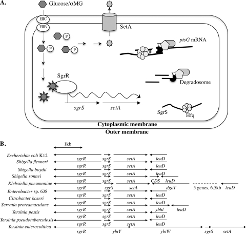
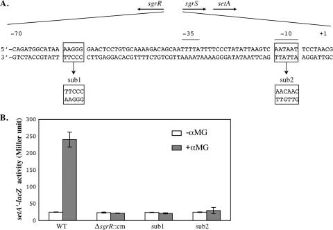
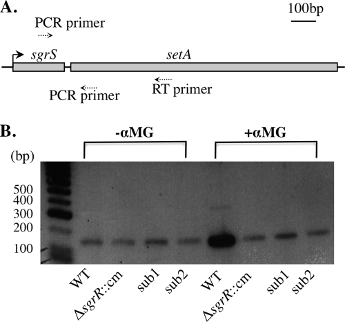
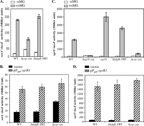
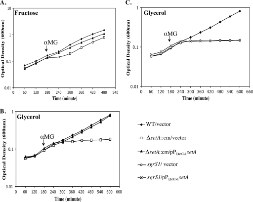
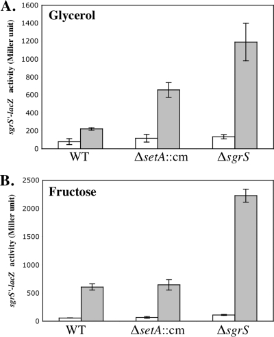
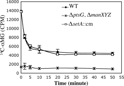
Accumulation of certain nonmetabolizable sugar-phosphates (including α-methyl glucoside-6-phosphate) in Escherichia coli is growth inhibitory and elicits the glucose-phosphate stress response. The transcription factor SgrR activates transcription of the small RNA SgrS under stress conditions. SgrS represses translation of mRNAs encoding sugar transporters. The sgrR and sgrS genes are located directly upstream of setA, and this gene organization is conserved in numerous enteric species, prompting the hypothesis that SetA contributes to the glucose-phosphate stress response. SetA is a proton motive force-driven efflux pump capable of transporting various sugars and sugar analogs in vitro. This study demonstrates that setA expression is induced in response to glucose-phosphate stress, and this requires SgrR. Under stress conditions, setA is cotranscribed with sgrS from the sgrS promoter. A setA mutant exhibits a growth defect under stress conditions that can be complemented by setA in trans, suggesting that SetA contributes to the optimal cellular recovery from stress. Despite previous in vitro evidence that SetA can promote efflux of the stress-causing glucose analog α-methyl glucoside, in vivo data in this study indicate that SetA is not the major efflux pump responsible for removal of α-methyl glucoside under stress conditions.


Figures

FIG. 1.
FIG. 1.
Involvement of SgrS and SetA in the glucose-phosphate stress response. (A) Model of glucose-phosphate stress response in E. coli. (B) Organization of genes in the sgrR-sgrS-setA chromosomal region in organisms related to E. coli K-12. The directions of gene transcription are indicated by the arrows.
FIG. 2.
FIG. 2.
SgrR-dependent activation of setA expression. (A) Genetic organization of the sgrR-sgrS region. The directions of transcription of the sgrR and sgrS genes are depicted by the arrows at the top. The −35 and −10 promoter elements are indicated by horizontal lines above the nucleotide sequence. The boxed nucleotides show the positions where mutations sub1 and sub2 were constructed. The substituted sequences are shown below the wild-type sequence. (B) Cells were grown in LB, and 0.1% αMG was added to half of the cultures. β-Galactosidase activity was assayed 45 min after the addition of αMG. The error bars indicate the standard deviations of three independent experiments.
FIG. 3.
FIG. 3.
RT-PCR analysis of sgrS and setA. (A) Schematic diagram showing the positions of the RT primer and PCR primers within the sgrS-setA region. (B) Wild-type (DJ480), ΔsgrR::cm mutant (CV700), and sgrS promoter mutant sub1 (YS135) and sub2 (YS137) cells were grown in LB medium at 37°C to an OD600 of ∼0.5. Total RNA was prepared from cells before and 10 min after exposure to 0.5% αMG, reverse transcribed, and amplified by PCR as described in Materials and Methods. Control experiments were also performed on each sample without addition of the reverse transcriptase, and no PCR product was detected in any samples (data not shown).
FIG. 4.
FIG. 4.
Effects of CRP and KdgR on setA and sgrS expression. (A) Cells were grown in LB to an OD600 of ∼0.5, and 0.1% αMG was added to half of the cultures. β-Galactosidase activity was assayed 45 min after the addition of αMG. (B) Cells were grown in LB supplemented with 100 μg/ml ampicillin to an OD600 of ∼0.5 and assayed for β-galactosidase activity. (C) Cells were grown in LB to an OD600 of ∼0.5, and 0.001% αMG was added to half of the cultures. β-Galactosidase activity was assayed 45 min after the addition of αMG. (D) Cells were grown in LB supplemented with 100 μg/ml ampicillin to an OD600 of ∼0.5 and assayed for β-galactosidase activity. The error bars indicate standard deviations from three independent experiments.
FIG. 5.
FIG. 5.
Growth of wild-type and ΔsetA strains in the presence of αMG. Cells were grown at 37°C in minimal MOPS medium supplemented with 1 ng/ml aTc, 25 μg/ml kanamycin, and 0.2% fructose (A) or 0.4% glycerol (B and C) to an OD600 of ∼0.1, and αMG was added to the cultures to a final concentration of 0.5%. Each graph is representative of three independent trials.
FIG. 6.
FIG. 6.
Effects of setA mutation on sgrS′-lacZ activity in response to αMG. Cells were grown in minimal MOPS medium supplemented with 0.4% glycerol (A) or 0.2% fructose (B), and 0.001% αMG was added to half of the cultures. β-Galactosidase activity was assayed after 45 min of αMG addition. White bars represent the absence of αMG, while gray bars represent the presence of αMG. The error bars indicate the standard deviations of three independent experiments.
FIG. 7.
FIG. 7.
Effects of setA mutation on the efflux of [14C]αMG. Wild-type (DJ480), ΔsetA::cm mutant (ST101), and ΔmanXYZ::kan ΔptsG::cm mutant (CV107) strains were grown at 37°C in minimal MOPS medium supplemented with 0.4% glycerol to an OD600 of ∼0.1. The cells were incubated with [14C]αMG (3.3 μM; 1 μCi/ml) at room temperature for 20 min and then diluted 200-fold with fresh minimal MOPS-glycerol medium. Radioactivity was examined at the indicated times as described in Materials and Methods. The error bars indicate the standard deviations of three independent experiments.

Similar articles

Cited by

References

    1. Baba, T., T. Ara, M. Hasegawa, Y. Takai, Y. Okumura, M. Baba, K. A. Datsenko, M. Tomita, B. L. Wanner, and H. Mori. 2006. Construction of Escherichia coli K-12 in-frame, single-gene knock-out mutants—the Keio collection. Mol. System Biol. doi:10.1038/msb4100050. - DOI - PMC - PubMed
    1. Bensing, B. A., B. J. Meyer, and G. M. Dunny. 1996. Sensitive detection of bacterial transcription initiation sites and differentiation from RNA processing sites in the pheromone-induced plasmid transfer system of Enterococcus faecalis. Proc. Natl. Acad. Sci. U. S. A. 93:7794-7799. - PMC - PubMed
    1. Clepet, C., I. L. Clainche, and M. Caboche. 2004. Improved full-length cDNA production based on RNA tagging by DNA ligase. Nucleic Acids Res. 32:e6. - PMC - PubMed
    1. Condemine, G. 2000. Characterization of SotA and SotB, two Erwinia chrysanthemi proteins which modify isopropyl-β-D-thiogalactopyranoside and lactose induction of the Escherichia coli lac promoter. J. Bacteriol. 182:1340-1345. - PMC - PubMed
    1. Dippel, R., and W. Boos. 2005. The maltodextrin system of Escherichia coli: metabolism and transport. J. Bacteriol. 187:8332-8339. - PMC - PubMed

Publication types

MeSH terms

LinkOut - more resources