. 2011 Jan;193(1):163-76.
doi: 10.1128/JB.00852-10. Epub 2010 Oct 22.

The Burkholderia cenocepacia LysR-type transcriptional regulator ShvR influences expression of quorum-sensing, protease, type II secretion, and afc genes

Affiliations

The Burkholderia cenocepacia LysR-type transcriptional regulator ShvR influences expression of quorum-sensing, protease, type II secretion, and afc genes

Eoin P O'Grady et al. J Bacteriol. 2011 Jan.

Abstract

Burkholderia cenocepacia is a significant opportunistic pathogen in individuals with cystic fibrosis. ShvR, a LysR-type transcriptional regulator, has previously been shown to influence colony morphology, biofilm formation, virulence in plant and animal infection models, and some quorum-sensing-dependent phenotypes. In the present study, it was shown that ShvR negatively regulates its own expression, as is typical for LysR-type regulators. The production of quorum-sensing signal molecules was detected earlier in growth in the shvR mutant than in the wild type, and ShvR repressed expression of the quorum-sensing regulatory genes cepIR and cciIR. Microarray analysis and transcriptional fusions revealed that ShvR regulated over 1,000 genes, including the zinc metalloproteases zmpA and zmpB. The shvR mutant displayed increased gene expression of the type II secretion system and significantly increased protease and lipase activities. Both ShvR and CepR influence expression of a 24-kb genomic region adjacent to shvR that includes the afcA and afcC operons, required for the production of an antifungal agent; however, the reduction in expression was substantially greater in the shvR mutant than in the cepR mutant. Only the shvR mutation resulted in reduced antifungal activity against Rhizoctonia solani. ShvR, but not CepR, was shown to directly regulate expression of the afcA and afcC promoters. In summary, ShvR was determined to have a significant influence on the expression of quorum-sensing, protease, lipase, type II secretion, and afc genes.


Figures
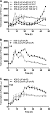
FIG. 1.
FIG. 1.
Influence of medium and temperature on shvR promoter activity. Expression of an shvR promoter::lux fusion (pPshvR) in K56-2 in LB and TSB media at 29°C or 37°C (A) and in K56-2 and K56-2ΔshvR in LB (B) and TSB (C) at 37°C. Expression was significantly increased in K56-2ΔshvR compared to that in K56-2 from 11 to 38 h in LB and from 22 to 35 h in TSB (P < 0.001, two-way ANOVA). All values are the means ± standard deviations (SD) of results from triplicate cultures and are representative of results from two individual trials.
FIG. 2.
FIG. 2.
Effect of the shvR mutation on AHL activity and expression of QS genes. (A) AHL activity was measured using the biosensor A. tumefaciens A136(pCF218)(pMV26) in coculture with B. cenocepacia at 29°C. AHL activity was significantly increased from 22 to 26.5 h (log phase) and significantly decreased from 35 to 45 h (stationary phase) in K56-2ΔshvR(pUCP28T) compared to that in K56-2(pUCP28T) (P < 0.05, two-way ANOVA). Expression of cepI (B), cepR (C), or cciIR (D) was monitored using promoter::lux fusions (pCP300, pRM432, or pRM445, respectively) in K56-2 and K56-2ΔshvR in LB at 37°C. Expression is significantly increased in K56-2ΔshvR compared to that in K56-2 from 24.5 to 40.5 h for cepI, from 5.5 to 49.5 h for cepR, and from 19 to 49.5 h for cciIR (P < 0.001, two-way ANOVA). All values are the means ± SD of results from triplicate cultures and are representative of results from at least two individual trials.
FIG. 3.
FIG. 3.
Regulation of biofilm formation and colony morphology. (A) Biofilm formation was assessed on polystyrene pegs. Statistical significance was determined (*, P < 0.001 [one-way ANOVA]). All values are the means ± SD of results from 8 replicate cultures and are representative of results from at least two individual trials. (B) Colony morphology of cultures spot inoculated in 8 replicates onto LB agar. Images are representative of results from two individual trials.
FIG. 4.
FIG. 4.
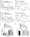
Expression of zmpA, zmpB, gspC, and the gspG operon and protease and lipase activities. Expression of zmpA (A), zmpB (B), gspC (D), or gspG (E) was monitored using promoter::lux fusions in B. cenocepacia in LB at 37°C. Expression is significantly decreased in K56-2ΔshvR compared to that in K56-2 from 20 to 37.5 h for zmpA and from 8 to 21.5 h for zmpB. Expression is significantly increased in K56-2ΔshvR compared to that in K56-2 from 16 to 32 h for gspC and gspG. Expression is significantly increased in K56-2ΔshvR including 300 pM OHL compared to that in K56-2ΔshvR from 17.5 to 26.5 h and 30.5 to 48 h for zmpA and 12 to 23.5 h and 32 to 40.5 h for zmpB (P < 0.001, two-way ANOVA). All values are the means ± SD of results from triplicate cultures and are representative of results from at least two individual trials. Cultures were spot inoculated in triplicate on D-BHI-1.5% skim agar for protease activity (C) or 1% Tween 80 agar for lipase activity (F), and zones of clearing or precipitation around colony growth were measured, respectively. Statistical significance was determined (*, P < 0.05; ***, P < 0.001 [one-way ANOVA]).
FIG. 5.
FIG. 5.
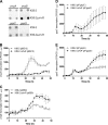
Expression of zmpA, zmpB, gspC, and the gspG operon on D-BHI-1.5% skim milk agar and in D-BHI-1.5% skim milk. (A) Expression of zmpA, zmpB, gspC, and gspG was monitored using promoter::lux fusions in B. cenocepacia from cultures spot inoculated onto D-BHI-1.5% skim milk agar. Images are representative of results from two individual trials. Expression of zmpA (B), zmpB (C), gspC (D), or gspG (E) was monitored using promoter::lux fusions in B. cenocepacia in D-BHI-1.5% skim milk at 37°C. Expression is significantly decreased in K56-2ΔshvR compared to that in K56-2 from 18 to 48 h for zmpA. Expression is significantly increased in K56-2ΔshvR compared to that in K56-2 from 29.5 to 48 h for zmpB, 38.5 to 48 h for gspC, and 35.5 to 48 h for gspG (P < 0.001, two-way ANOVA). All values are the means ± SD of results from triplicate cultures and are representative of results from at least two individual trials.
FIG. 6.
FIG. 6.
Genomic organization and expression of genes in the shvR/afc genomic region. (A) ShvR lies in the genomic region adjacent to the afcA and afcC operons, which are transcribed divergently. (B) Expression was measured in K56-2shvR::Tp (open bars) and K56-R2 (closed bars) compared to that of K56-2 as determined by microarray analysis. Changes in expression of BCAS0204 and BCAS0205 were included, as they are predicted to be part of the afcA operon (60), although they did not meet statistical significance for microarray analysis. Expression of afcA (C) and afcC (D) was monitored using promoter::lux fusions in B. cenocepacia in LB at 37°C. Expression is significantly decreased in K56-2ΔshvR versus K56-2 (P < 0.001, two-way ANOVA). All values are the means ± SD of results from triplicate cultures and are representative of results from at least two individual trials.
FIG. 7.
FIG. 7.
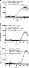
Expression of cepI, afcA, and afcC in the heterologous host E. coli by using an expression-reporter system. Strains harbor two plasmids: a derivative of pUCP28T and a promoter::lux fusion for cepI (pEPO100), afcA (pEPO101), or afcC (pEPO128) as indicated. Assays were performed in the absence or presence of 3,000 pM OHL at 37°C. Expression of cepI (A), afcA (B), or afcC (C) was monitored using the appropriate promoter::lux fusion in E. coli also carrying pUCP28T, or cepR in trans (pSLR100) or shvR in trans (p28T-shvR). All values are the means ± SD of results from triplicate cultures and are representative of results from at least two individual trials.
FIG. 8.
FIG. 8.
Antifungal activity against R. solani. R. solani was grown on malt agar in the presence of K56-2, K56-R2 (cepR), K56-2ΔshvR(pUCP28T), or K56-2ΔshvR(p28T-shvR). Fungal growth inhibition was recorded after 3 days. The assay was performed in triplicate; a representative plate for each assay is shown.

Similar articles

Cited by

References

    1. Ausubel, F., R. Brent, R. Kingston, D. Moore, J. Seidman, J. Smith, and K. Struhl. 1989. Current protocols in molecular biology, vol. 1. John Wiley & Sons, Inc., New York, NY.
    1. Baldwin, A., P. A. Sokol, J. Parkhill, and E. Mahenthiralingam. 2004. The Burkholderia cepacia epidemic strain marker is part of a novel genomic island encoding both virulence and metabolism-associated genes in Burkholderia cenocepacia. Infect. Immun. 72:1537-1547. - PMC - PubMed
    1. Bernier, S. P., A. L. Beeston, and P. A. Sokol. 2008. Detection of N-acyl homoserine lactones using a traI-luxCDABE-based biosensor as a high-throughput screening tool. BMC Biotechnol. 8:59. - PMC - PubMed
    1. Bernier, S. P., D. T. Nguyen, and P. A. Sokol. 2008. A LysR-type transcriptional regulator in Burkholderia cenocepacia influences colony morphology and virulence. Infect. Immun. 76:38-47. - PMC - PubMed
    1. Bernier, S. P., and P. A. Sokol. 2005. Use of suppression-subtractive hybridization to identify genes in the Burkholderia cepacia complex that are unique to Burkholderia cenocepacia. J. Bacteriol. 187:5278-5291. - PMC - PubMed

Publication types

LinkOut - more resources