. 2011 Jan;193(1):63-74.
doi: 10.1128/JB.00839-10. Epub 2010 Oct 22.

The Rcs signal transduction pathway is triggered by enterobacterial common antigen structure alterations in Serratia marcescens

Affiliations

The Rcs signal transduction pathway is triggered by enterobacterial common antigen structure alterations in Serratia marcescens

María E Castelli et al. J Bacteriol. 2011 Jan.

Abstract

The enterobacterial common antigen (ECA) is a highly conserved exopolysaccharide in Gram-negative bacteria whose role remains largely uncharacterized. In a previous work, we have demonstrated that disrupting the integrity of the ECA biosynthetic pathway imposed severe deficiencies to the Serratia marcescens motile (swimming and swarming) capacity. In this work, we show that alterations in the ECA structure activate the Rcs phosphorelay, which results in the repression of the flagellar biogenesis regulatory cascade. In addition, a detailed analysis of wec cluster mutant strains, which provoke the disruption of the ECA biosynthesis at different levels of the pathway, suggests that the absence of the periplasmic ECA cyclic structure could constitute a potential signal detected by the RcsF-RcsCDB phosphorelay. We also identify SMA1167 as a member of the S. marcescens Rcs regulon and show that high osmolarity induces Rcs activity in this bacterium. These results provide a new perspective from which to understand the phylogenetic conservation of ECA among enterobacteria and the basis for the virulence attenuation detected in wec mutant strains in other pathogenic bacteria.


Figures

FIG. 1.
FIG. 1.
(A) Flagellin immunodetection. Whole-cell extracts from cells grown overnight in LB medium at 37°C were analyzed by Western blotting, in which the blots were developed with antiflagellin polyclonal antibodies. (B) Zymogram analysis. The spent culture medium from strains grown overnight in LB medium at 37°C was analyzed. Samples were prepared and standardized as described in Materials and Methods. Phospholipase activity is visualized as a white precipitate band that comigrates with the 26-kDa marker. (C) Motility assays. Swimming (LB medium, 0.25% agar) plates were incubated overnight at 37°C (upper panel), and swarming (LB medium, 0.6% agar) plates were incubated overnight at 30°C (lower panel). Results are representative of those from three individual assays with identical results. The following strains were analyzed: S. marcescens RM66262 (wild type [wt]); derived pKnock-Cm wecG mutant; complemented wecG mutant (wecG/pYG); and double mutants 7695, 7696, and 7697. (D) Schematic representation of the rcs genes in Serratia marcescens. The sites of the mini-Tn5 Km transposon insertions in strains 7695, 7696, and 7697 are indicated with arrows.
FIG. 2.
FIG. 2.
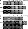
Motility assays. Swimming (LB medium, 0.25% agar) plates (A) and swarming (LB medium, 0.6% agar) plates (B) were incubated overnight at 37°C and 30°C, respectively. The following strains were analyzed: S. marcescens RM66262 (wild type [wt]); derived pKnock-Gm rcsB, rcsA, rcsC, and rcsF insertion mutants; mini-Tn5 Km mutants wecD and wzyE; complemented wzyE mutant (wzyE/pYG); and double mutants wecD rcsA, wecD rcsB, wecD rcsC, wecD rcsF, wzyE rcsA, wzyE rcsB, wzyE rcsC, and wzyE rcsF. Results are representative of those from three individual assays with identical results.
FIG. 3.
FIG. 3.
Flagellin immunodetection. Whole-cell extracts from cultures of the indicated S. marcescens strains grown overnight in liquid LB medium (swimming) (A) or obtained from the edge of LB medium-0.6% agar plates (swarming) (B) were analyzed by Western blotting in which the blots were developed with antiflagellin polyclonal antibodies. Samples were standardized as described in Materials and Methods. (C) Densitometric analysis of the flagellin immunodetections shown in panels A and B. Results were calculated from three independent immunodetection assays and are expressed in units relative to the value determined for the wild-type (wt) strain (swimming). Error bars indicate standard deviations.
FIG. 4.
FIG. 4.
(A) Quantitative RT-PCR analysis of flhD and SMA1167 expression. Total RNA was extracted from the indicated strains grown to mid-exponential phase in LB medium or LB medium-0.45 M NaCl (indicated NaCl) at 37°C. flhD and SMA1167 expression was analyzed by real-time RT-PCR using specific primers (Table 1), and 16S rRNA was used as the internal control. Average relative expression and fold change in gene expression in each mutant strain compared with the expression of the wild-type (wt) strain grown in LB medium were calculated from triplicate samples as described in Materials and Methods. Error bars indicate standard deviations. Note the different scales for the y axes. (B) Predicted RcsB-binding motif in the promoter regions of flhD and SMA1167. The consensus RcsB motif is compared to the region detected in S. marcescens SMA1167 promoter. (Lower panel) Alignment of E. coli, S. enterica, and S. marcescens flhD promoters. In E. coli the identified RcsB-binding site is shaded in gray, the transcription initiation site is indicated with an arrow, and the corresponding −35 and −10 regions are underlined. In S. enterica and S. marcescens, these putative elements are also indicated to highlight the high degree of conservation with the E. coli sequence.
FIG. 5.
FIG. 5.
Quantitative RT-PCR analysis of flhD (A) and SMA1167 (B) expression under swimming and swarming conditions. Total RNA was extracted from the indicated strains grown to mid-exponential phase in liquid LB medium (swimming) or obtained from the edge of LB medium-0.6% agar plates (swarming). flhD and SMA1167 expression was analyzed by real-time RT-PCR using specific primers (Table 1), and 16S rRNA was used as the internal control. Average relative expression and fold change in gene expression in each mutant strain compared with the expression of the wild-type (wt) strain grown in LB medium were calculated from triplicate samples as described in Materials and Methods. Error bars indicate standard deviations. Note the different scales for the y axes.
FIG. 6.
FIG. 6.
Biosynthetic pathway for the assembly of ECA. (A) Schematic representation of the wec cluster in S. marcescens; (B) enzymatic reactions and enzymes involved in the biosynthesis of ECA. Abbreviations: Und-P, undecaprenyl monophosphate; Und-PP, undecaprenyl pyrophosphate; TTP, dTTP; TDP, dTDP; PPi, inorganic pyrophosphate; Glc-1-P, glucose-1-phosphate; α-keto-glu, α-ketoglutarate; 4-keto-6-deoxy-d-Glc, 4-keto-6-deoxy-d-glucose, acetyl-CoA, acetyl coenzyme A; CoASH, coenzyme A; GlcNAc, N-acetylglucosamine; ManNAc, N-acetylmannose; ManNAcA, N-acetyl-d-mannosaminuronic acid; FucN, fucosamine; Fuc4NAc, 4-acetamido-4,6-dideoxy-d-galactose.
FIG. 7.
FIG. 7.
Phenotypic analysis of S. marcescens wecA and wzzE mutant strains. Exopolysaccharide preparations of the wild-type (wt) and mutant strains were analyzed by immunodetection with anti-O14 antiserum to detect ECA (A) or by silver staining to examine LPS (B). The localization of the high-molecular-weight bands pattern in ECA (*) and the core and O antigen in LPS are indicated. (C) Flagellin immunodetection. Whole-cell extracts from cultures of the indicated strains grown in LB medium overnight at 37°C were analyzed by Western blotting, in which the blots were developed with antiflagellin polyclonal antibodies. Loaded samples were standardized as described in Materials and Methods. (D) Quantitative RT-PCR analysis of flhD and SMA1167 expression. Total RNA was extracted from the indicated strains grown to mid-exponential phase in LB medium at 37°C. flhD and SMA1167 expression was analyzed by real-time RT-PCR using specific primers (Table 1), and 16S rRNA was used as the internal control. Average relative expression and fold change in gene expression in each mutant strain compared with the expression of the wild-type strain grown in LB were calculated from triplicate samples as described in Materials and Methods. Error bars indicate standard deviations. Note the different scales for the y axes.

Similar articles

Cited by

References

    1. Abramoff, M. D., P. J. Magelhaes, and S. J. Ram. 2004. Image processing with ImageJ. Biophotonics Int. 11:36-42.
    1. Alexander, D. C., and M. A. Valvano. 1994. Role of the rfe gene in the biosynthesis of the Escherichia coli O7-specific lipopolysaccharide and other O-specific polysaccharides containing N-acetylglucosamine. J. Bacteriol. 176:7079-7084. - PMC - PubMed
    1. Alexeyev, M. F. 1999. The pKNOCK series of broad-host-range mobilizable suicide vectors for gene knockout and targeted DNA insertion into the chromosome of gram-negative bacteria. Biotechniques 26:824-826, 828. - PubMed
    1. Arakawa, Y., R. Wacharotayankun, T. Nagatsuka, H. Ito, N. Kato, and M. Ohta. 1995. Genomic organization of the Klebsiella pneumoniae cps region responsible for serotype K2 capsular polysaccharide synthesis in the virulent strain Chedid. J. Bacteriol. 177:1788-1796. - PMC - PubMed
    1. Bahrani-Mougeot, F. K., E. L. Buckles, C. V. Lockatell, J. R. Hebel, D. E. Johnson, C. M. Tang, and M. S. Donnenberg. 2002. Type 1 fimbriae and extracellular polysaccharides are preeminent uropathogenic Escherichia coli virulence determinants in the murine urinary tract. Mol. Microbiol. 45:1079-1093. - PubMed

Publication types