Review
. 2011 Apr;32(2):159-224.
doi: 10.1210/er.2009-0039. Epub 2010 Oct 22.

Child health, developmental plasticity, and epigenetic programming

Affiliations
Review

Child health, developmental plasticity, and epigenetic programming

Z Hochberg et al. Endocr Rev. 2011 Apr.

Abstract

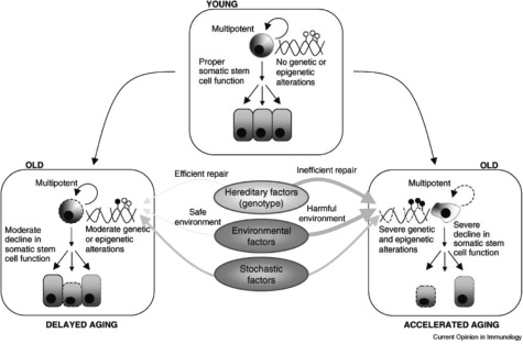
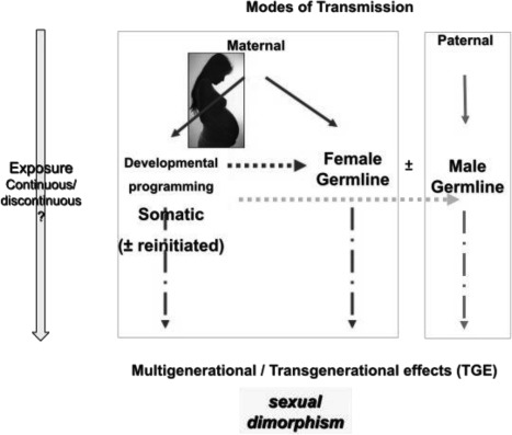
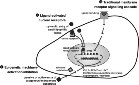
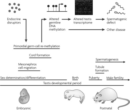
Plasticity in developmental programming has evolved in order to provide the best chances of survival and reproductive success to the organism under changing environments. Environmental conditions that are experienced in early life can profoundly influence human biology and long-term health. Developmental origins of health and disease and life-history transitions are purported to use placental, nutritional, and endocrine cues for setting long-term biological, mental, and behavioral strategies in response to local ecological and/or social conditions. The window of developmental plasticity extends from preconception to early childhood and involves epigenetic responses to environmental changes, which exert their effects during life-history phase transitions. These epigenetic responses influence development, cell- and tissue-specific gene expression, and sexual dimorphism, and, in exceptional cases, could be transmitted transgenerationally. Translational epigenetic research in child health is a reiterative process that ranges from research in the basic sciences, preclinical research, and pediatric clinical research. Identifying the epigenetic consequences of fetal programming creates potential applications in clinical practice: the development of epigenetic biomarkers for early diagnosis of disease, the ability to identify susceptible individuals at risk for adult diseases, and the development of novel preventive and curative measures that are based on diet and/or novel epigenetic drugs.


Figures

Fig. 1.
Fig. 1.
Preadult periods of adaptive plasticity in the transition between life-history phases (double arrows). Prenatal growth affects adult health and disease. The transition from infancy to childhood confers a predictive adaptive response that determines adult height. The transition from childhood to juvenility bestows an adaptive response that resolves adult body composition and metabolic consequences. The transition from juvenility to adolescence establishes longevity and the age of reproduction and fecundity. IC, Infancy-childhood (transition).
Fig. 2.
Fig. 2.
The match-mismatch paradigm of metabolic disease. The developing organism senses maternally transmitted environmental cues, such as undernutrition, during prenatal and early postnatal life. Developmental plasticity in response to these cues modifies the default trajectory defined by the inherited fetal genome and epigenome according to whether the environment is perceived as adequate (dark background) or deprived (light background), resulting in adjustment of metabolic set points. If the eventual mature environment, whether adequate or deprived, matches the prediction, then the risk of metabolic disease in later life is low. If there is a mismatch between the predicted and actual mature environments, particularly if the mature environment is richer than anticipated, then the risk of metabolic disease is enhanced. [Reproduced from P. D. Gluckman et al.: Am J Hum Biol 19:1–19, 2007 (23). © 2006 Wiley-Liss, Inc.; reprinted with permission from John Wiley & Sons, Inc.]
Fig. 3.
Fig. 3.
Top panel, DNA vs. chromatin. The genome is the invariant DNA sequence of an individual. The epigenome is the overall chromatin composition, which indexes the entire genome in any given cell. It varies according to cell type and response to the internal and external signals that it receives. Lower panel, Epigenome diversification occurs during development in multicellular organisms as differentiation proceeds from a single stem cell (the fertilized embryo) to more committed cells. Reversal of differentiation or transdifferentiation requires the reprogramming of the cell's epigenome. [Fig. 3 and its legend have been reproduced with permission from C. D. Allis et al.: Epigenetics, Chap 3, Cold Spring Harbor Laboratory Press, Woodbury, NY, 2007 (561). © 2007 CSHL Press.]
Fig. 4.
Fig. 4.
Alterations in methylation status during development. During embryonic development and gonadal sex determination, primordial germ cells undergo genome-wide demethylation, which erases previous parental-specific methylation marks that regulate imprinted gene expression. In the male germ line, paternal methylation marks in imprinted genes are laid down in developing gonocytes that will develop into spermatogonia. The female germ line establishes maternal methylation marks in imprinted genes at a later stage. After fertilization, the paternal genome is actively demethylated, whereas the maternal genome undergoes passive demethylation (176). Genome-wide remethylation occurs on both parental genomes before implantation. However, imprinted genes maintain their methylation marks throughout this reprogramming, allowing for the inheritance of parental-specific monoallelic expression in somatic tissues throughout adulthood. [Reprinted with permission from R. L. Jirtle and M. K. Skinner: Nat Rev Genet 8:253–262, 2007 (62). © 2007 Macmillan Publishers Ltd.]
Fig. 5.
Fig. 5.
Histone modifications can generate both short-term and long-term outcomes. The amino-terminal tails of all eight core histones protrude through the DNA and are exposed on the nucleosome surface, where they are subject to an enormous range of enzyme-catalyzed modifications of specific amino-acid side chains, including acetylation of lysines, methylation of lysines and arginines, and phosphorylation of serines and threonines. Histone tail modifications are put in place by modifying and demodifying enzymes whose activities can be modulated by environmental and intrinsic signals. Modifications may function in short-term, ongoing processes (such as transcription, DNA replication, and repair) and in more long-term functions (as determinants of chromatin conformation, for example heterochromatin formation, or as heritable markers that both predict and are necessary for future changes in transcription). Short-term modifications are transient and show rapidly fluctuating levels. Long-term, heritable modifications need not necessarily be static; in theory, they still show enzyme-catalyzed turnover, but the steady-state level must be relatively consistent. [Reprinted with permission from B. M. Turner: Nat Cell Biol 9:2–6, 2007 (562). © 2007 Macmillan Publishers Ltd.]
Fig. 6.
Fig. 6.
The epigenotype model of developmental origins of disease. Environmental factors acting in early life have consequences that become manifest as an altered disease risk in later life. The period of life in which external factors can influence biology extends from conception to the neonatal period and early infancy. It has been suggested that the baby receives from its mother a forecast of the environment it will encounter after birth and modifies its metabolism, whole body physiology, and growth trajectory appropriately to maximize its chances of survival postnatally. However, these adaptations become detrimental if the conditions after birth are not the same as the ones encountered during early life. These adaptations include metabolic and endocrine changes that may lead to lifelong changes in the function and structure of the body—a concept that has been termed programming. The molecular mechanisms by which a phenomenon that occurs in utero has a phenotypic consequence many years later are likely to involve epigenetic mechanisms of gene regulation. Epigenetic marks can be modulated by environmental factors, are heritable, and perpetuate gene-expression changes that underlie programming and may contribute to the onset of disease in later life. Ac, Histone acetylation/active genes; CH3, DNA methylation/silent genes. [Reprinted with permission from I. Sandovici et al.: Epigenetics, Horizon Scientific Press/Caister Academic Press, Norfolk, UK, 2008 (563). © with permission from the publisher]
Fig. 7.
Fig. 7.
The two imprinted domains of the 11p15 chromosomal region are under the control of two ICRs. The reciprocal imprinting of the maternally (mat) expressed H19 and the paternally (pat) expressed IGF2 depends on an ICR1 located upstream from the H19 gene that acts as an insulator. The repressor factor CTCF (CCCTC-binding factor) binds to the unmethylated maternal copy of the ICR and prevents the IGF2 gene promoter from interacting with enhancers downstream from the H19 gene. This results in transcriptional silencing of the maternal IGF2 allele. On the paternal allele, the ICR is methylated, and CTCF binding is prevented. This leads to IGF2 transcription on the paternal allele and silencing of the H19 gene. The centromeric KCNQ1 domain produces a noncoding RNA (antisense KCNQ1OT1 RNA) that silences many of the genes in this domain. Paternally expressed genes are represented as white boxes, maternally expressed genes as black boxes, and nonexpressed genes as gray boxes. BWS is associated with a variety of genetic and epigenetic defects within the imprinted 11p15 region. Most patients (70%) exhibit an epigenetic defect. Ten percent of BWS patients display an imprinting defect at the IGF2-H19 domain (aberrant GOM at the maternal copy of the ICR), which results in silencing of the maternal H19 gene and a biallelic expression of the IGF2 gene. The majority of the BWS patients exhibit a LOM at the ICR of the KCNQ1 domain. Loss of methylation at this ICR results in activation of the normally silent maternal allele of KCNQ1OT1 and CDKN1C silencing. In SRS, the mirror phenotype of BWS, a loss of imprinting at the IGF2–H19 domain was identified: the paternal allele switches to a maternal epigenotype, and this results in biallelic expression of H19 and loss of IGF2 expression. Genetic and environmental factors could induce these epigenetic anomalies.
Fig. 8.
Fig. 8.
Potential mechanisms for environmental influences on developmental establishment of DNA methylation. A, Nutritional or other stimuli that affect either the efficiency of one-carbon metabolism or the activity of DNMT1 could alter the developmental establishment of DNA methylation at metastable epialleles. Flux through the transmethylation/remethylation pathway is dependent upon nutrients including folate, vitamins B12 and B6, choline, betaine, and methionine. B, Transcriptional activity during critical developmental periods can impair de novo methylation. Any nutritional or other environmental exposure that activates gene transcription during periods of de novo CpG methylation can permanently imprint transcriptional competence by preventing hypermethylation. Methylated CpG sites are shown as “filled lollipops.” Although a gene promoter region is shown here, similar effects could occur at any genomic region contributing to transcriptional regulation, such as a distal enhancer. 5CH3THF, 5-Methyl tetrahydrofolate; SAH, S-adenosylhomocysteine; DMG, dimethyl glycine. [Reprinted with permission from R. A. Waterland and K. B. Michels: Annu Rev Nutr 27:363–388, 2007 (55). © Annual Reviews.]
Fig. 9.
Fig. 9.
Parental imprints are established during oogenesis or spermatogenesis at sequence elements that control the imprinted expression (the ICRs). After fertilization of the egg by the sperm, these imprints are maintained throughout development. DNA methylation (lollipops) is the most consistent hallmark of imprints. Two examples of ICRs are depicted: one with paternally derived (ICR1) and one with maternally derived (ICR2) DNA methylation. [Reprinted from K. Delaval and R. Feil: Curr Opin Genet Dev 14:188–195, 2004 (52), with permission from Elsevier.]
Fig. 10.
Fig. 10.
A model of how genetic and epigenetic factors can affect aging. Young adult stem cells present no alterations in either the genetic or epigenetic levels, and so there is proper stem cell function and, consequently, tissue regeneration. Genotypes of low efficiency in repairing genetic or epigenetic (represented as lollipops over the structure of the DNA) defects or in maintaining epigenetic stability accompanied by harmful environmental exposures can accelerate the accumulation of molecular alterations at the genetic and the epigenetic levels, which in turn can accelerate the aging process. On the other hand, genotypes that are highly efficient in repairing genetic and epigenetic defects and in maintaining epigenetic stability accompanied by harmless environmental exposures can slow the accumulation of molecular alterations at the genetic and epigenetic levels, which, in turn, can delay the aging process. [Reprinted from F. M. Fraga: Curr Opin Immunol 21:446–453, 2009 (185). © with permission from Elsevier.]
Fig. 11.
Fig. 11.
Epigenetic regulation of sex-specific CYPs. Female-specific Cyp genes are proposed to be repressed in the male liver, and male-specific Cyp genes are proposed to be repressed in the female liver by packaging in heterochromatin. Continuous GH is proposed to activate female-specific genes, such as Cyp3a genes, by a mechanism that involves the local conversion of heterochromatin to euchromatin, which enables the binding of transcription factors (TF) that activate CYP gene expression. This process could involve the loss of DNA CpG methylation and/or loss of chromatin marks that are associated with repressed chromatin, such as histone H3 lysine 27 trimethylation, which is typically found in genes in a compact chromatin structure and is associated with a stable, inactive heterochromatic state. [Reprinted from D. J. Waxman and M. G. Holloway: Mol Pharmacol 76:215–228, 2009 (389), with permission from the publisher.]
Fig. 12.
Fig. 12.
Sexual dimorphism in the modes of transmission and its effects on the offspring in successive generations. The sex specificity of these effects operates at three different levels: 1) the maternal transmission during pregnancy and postnatal periods; 2) the sex of the parent who transmits the consequences of a stimulus exposure via the germline; and 3) the sex of the offspring who displays the maternal effect or paternal and/or maternal germline TGEs. [Reprinted from A. Gabory et al.: Mol Cell Endocrinol 304:8–18, 2009 (378), with permission from Elsevier.]
Fig. 13.
Fig. 13.
The three signaling pathways transduce environmental signals from the cell membrane to the chromatin structure in epigenetic programming of the genome: 1) activation or inhibition of the chromatin epigenetic machinery by metabolites of these substrates; 2) activation of nuclear receptors by ligands; and 3) traditional membrane receptor signaling cascades. [Reprinted from A. Gabory et al.: Mol Cell Endocrinol 304:8–18, 2009 (378), with permission from Elsevier.]
Fig. 14.
Fig. 14.
A model for endocrine-disruptor-induced epigenetic transgenerational disease. Endocrine-disruptor action reprograms the epigenome of the developing germ cell during embryonic sex determination, leading to genes and other DNA sequences with altered DNA methylation. These changes are proposed to alter the transcriptomes of the testis and other organs, thereby promoting adult pathologies, some of which are inherited transgenerationally. Epigenetic mechanisms might therefore have a role in the induction of adult-onset disease through environmental exposures early in development. [Reprinted with permission from R. L. Jirtle and M. K. Skinner: Nat Rev Genet 8:253–262, 2007 (62). © 2007 Macmillan Publishers Ltd.]

References

    1. Bateson P. 2005. The return of the whole organism. J Biosci 30:31–39 - PubMed
    1. Crews D, McLachlan JA. 2006. Epigenetics, evolution, endocrine disruption, health, and disease. Endocrinology 147:S4–S10 - PubMed
    1. Hochberg Z. 2009. Evo-devo of child growth II: human life history and transition between its phases. Eur J Endocrinol 160:135–141 - PubMed
    1. Barker DJ. 1995. Fetal origins of coronary heart disease. BMJ 311:171–174 - PMC - PubMed
    1. Barker DJ. 1992. The fetal origins of adult hypertension. J Hypertens 10:S39–S44 - PubMed

Publication types