. 2011 Jan;89(1):57-62.
doi: 10.1189/jlb.0610381. Epub 2010 Oct 22.

Store-operated Ca²+ signaling in dendritic cells occurs independently of STIM1

Affiliations

Store-operated Ca²+ signaling in dendritic cells occurs independently of STIM1

Bidhan C Bandyopadhyay et al. J Leukoc Biol. 2011 Jan.

Abstract

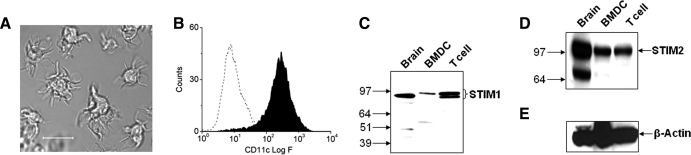
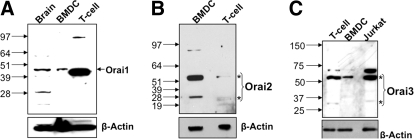
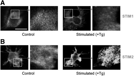
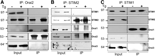
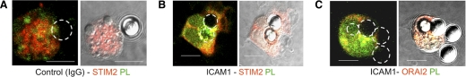
SOCE via CRAC channels is a critical signaling event in immune cells. Recent studies have identified key proteins underlying this process; STIM is an ER Ca²+ sensor that interacts with Orai, an intrinsic, pore-forming protein of the CRAC channel. In heterologous expression systems, STIM1 regulates SOCE by interacting with Orai1, -2, and -3. In native tissues, however, the precise roles of STIM and Orai proteins are not well defined. Here, we have investigated the molecular components of SOCE signaling in mouse DCs. We show that DCs predominantly express STIM2 and only very low levels of STIM1 compared with T lymphocytes. Upon store depletion with Tg, STIM2 aggregates and interacts selectively with Orai2. In contrast, Tg fails to aggregate STIM1 or enhance STIM1-mediated interactions with Orai proteins. Consistent with this biochemical characterization, stimulation of DCs with the adhesion molecule ICAM-1 selectively recruits STIM2 and Orai2 to the IS. Together, these data demonstrate a novel, STIM2-dependent SOCE signaling pathway in DCs.


Figures

Figure 1.
Figure 1.. Expression of STIM proteins in murine DCs.
(A) Bright-field image of BM-derived DCs with typical dendritic processes (original scale bar, 20 μm). (B) Flow cytometric analysis shows that enriched cultures were >90% positive for the DC marker, CD11c. (C) Western blots showing the expression of STIM1 and (D) STIM2 in DCs. Whole cell lysates (30 μg) were used for each sample. Whole mouse brain and mouse T cells were used as positive controls. (E) The blots were stripped and reprobed with β-actin as a loading control. BMDC, BM-derived DC.
Figure 2.
Figure 2.. Expression of Orai proteins in DCs.
Western blots showing the expression of (A) Orai1, (B) Orai2, and (C) Orai3 in DCs and T cells. Whole cell lysates (30 μg) were used for each sample. Whole mouse brain, T cells, and Jurkat T cells were used as positive controls. The blots were stripped and reprobed with β-actin as a loading control. *Monomer and dimer of Orai proteins.
Figure 3.
Figure 3.. Tg induces aggregation of STIM2.
(A) Immunofluorescence labeling of DCs for (A) STIM1 and (B) STIM2 under control conditions (unstimulated) and after Tg treatment (Stimulated; 2 μM Tg for 10 min). Fixed cells were permeabilized and stained with STIM1 and STIM2 antibody followed by a secondary antibody (FITC-conjugated), which alone, produced negligible staining (see Supplemental Fig. 2B).
Figure 4.
Figure 4.. Tg induces the association of STIM2 and Orai2 but not STIM1.
(A and B) Coimmunoprecipitation showing the enhanced association between STIM2 with Orai2 upon Tg treatment. Although detected in the immunoprecipitation (IP) complex, Orai1 levels are not increased by Tg treatment. (C) STIM2 and Orai2 are not present in the coimmunoprecipitation using a STIM1 antibody. Moreover, Tg treatment does not alter levels of Orai1 in this immunoprecipitation complex.
Figure 5.
Figure 5.. Orai2 and STIM2 are localized at the IS.
Representative confocal images of STIM2 and ORAI2 immunostaining in DCs stimulated with IgG (A)- or ICAM-1 (B and C)-coated beads. Circles indicate positions of beads clustered with DCs (original scale bars, 15 μm). Cells were colabeled with F-actin [phalloidin (PL), green] to reveal actin polarization toward contact sites. STIM2 (red) and ORAI2 (red) are polarized along with F-actin to the ICAM1-coated bead, as evident by the merged (yellow) staining. Immunostaining was assessed by a blinded scoring of >40 random conjugates from three independent experiments.

Similar articles

Cited by

References

    1. Hsu S.f., O'Connell P. J., Klyachko V. A., Badminton M. N., Thomson A. W., Jackson M. B., Clapham D. E., Ahern G. P. (2001) Fundamental Ca2+ signaling mechanisms in mouse dendritic cells: CRAC is the major Ca2+ entry pathway. J. Immunol. 166, 6126–6133 - PubMed
    1. Czerniecki B. J., Carter C., Rivoltini L., Koski G. K., Kim H. I., Weng D. E., Roros J. G., Hijazi Y. M., Xu S., Rosenberg S. A., Cohen P. A. (1997) Calcium ionophore-treated peripheral blood monocytes and dendritic cells rapidly display characteristics of activated dendritic cells. J. Immunol. 159, 3823–3837 - PubMed
    1. Koski G. K., Schwartz G. N., Weng D. E., Czerniecki B. J., Carter C., Gress R. E., Cohen P. A. (1999) Calcium mobilization in human myeloid cells results in acquisition of individual dendritic cell-like characteristics through discrete signaling pathways. J. Immunol. 163, 82–92 - PubMed
    1. Liou J., Kim M. L., Heo W. D., Jones J. T., Myers J. W., Ferrell J. E., Jr., Meyer T. (2005) STIM is a Ca2+ sensor essential for Ca2+-store-depletion-triggered Ca2+ influx. Curr. Biol. 15, 1235–1241 - PMC - PubMed
    1. Roos J., DiGregorio P. J., Yeromin A. V., Ohlsen K., Lioudyno M., Zhang S., Safrina O., Kozak J. A., Wagner S. L., Cahalan M. D., Velicelebi G., Stauderman K. A. (2005) STIM1, an essential and conserved component of store-operated Ca2+ channel function. J. Cell Biol. 169, 435–445 - PMC - PubMed

Publication types

MeSH terms