. 2010 Dec 1;185(11):6535-44.
doi: 10.4049/jimmunol.1002009. Epub 2010 Oct 22.

Cyclooxygenase-2 enhances antimicrobial peptide expression and killing of Staphylococcus aureus

Affiliations

Cyclooxygenase-2 enhances antimicrobial peptide expression and killing of Staphylococcus aureus

Jamie J Bernard et al. J Immunol. .

Abstract

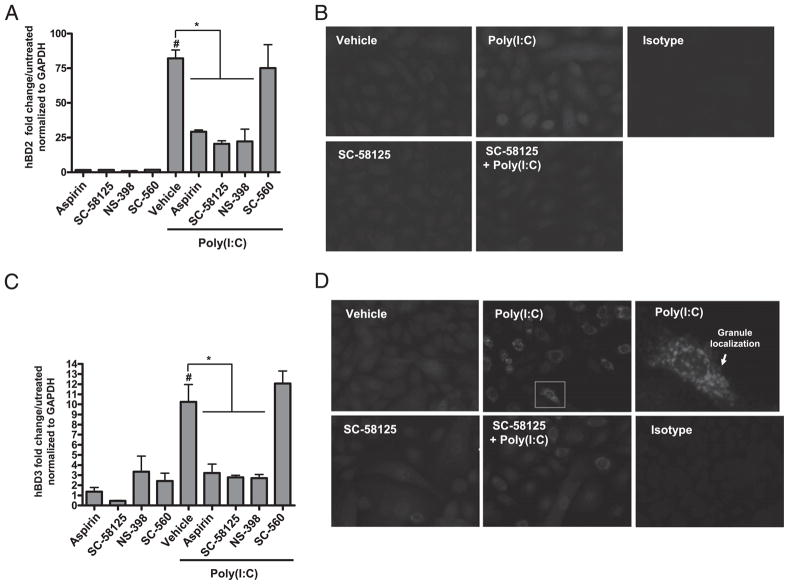
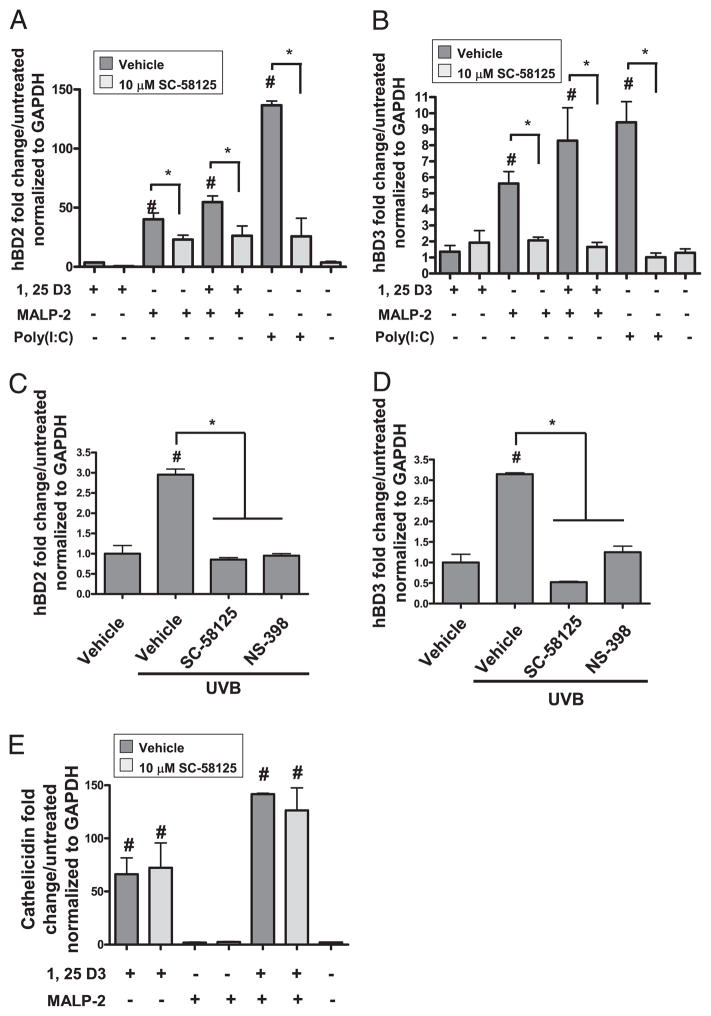
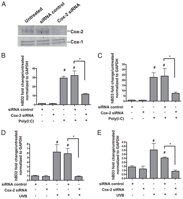
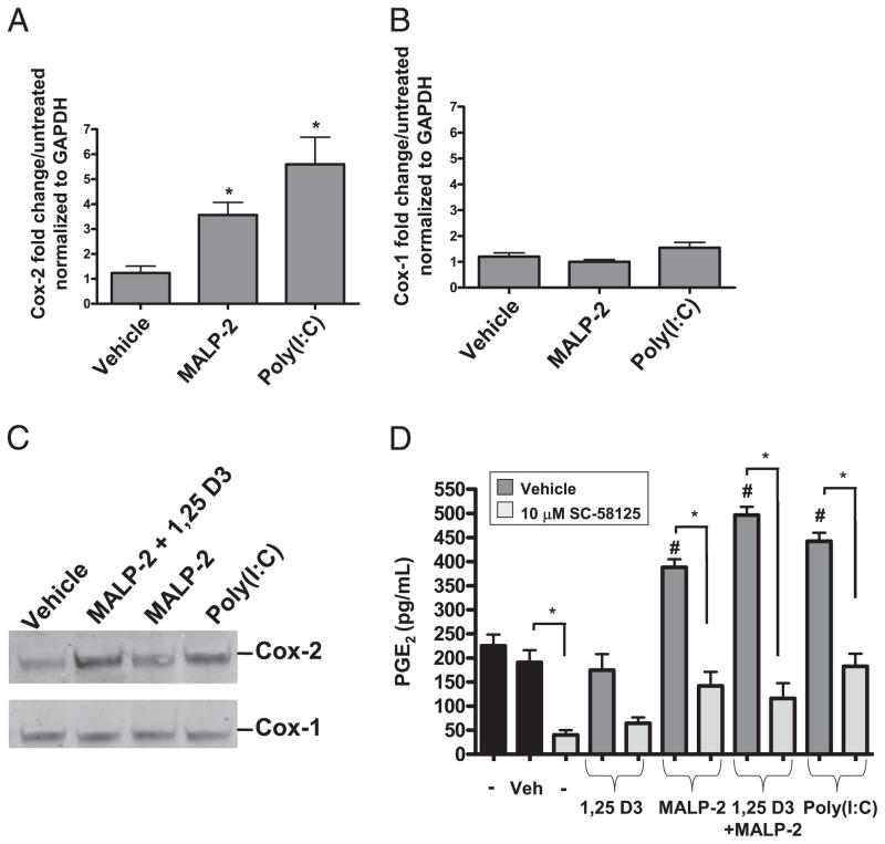
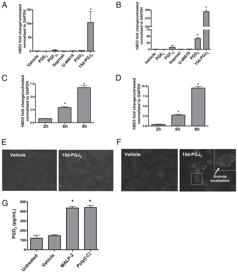
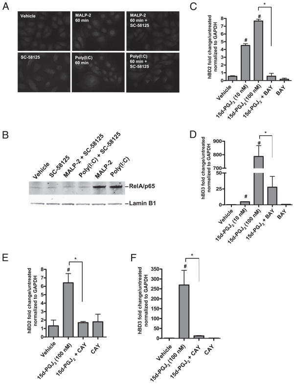
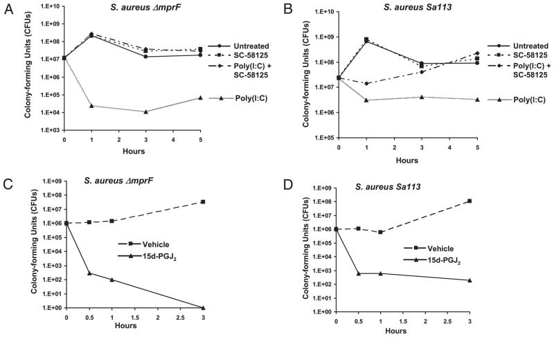
Antimicrobial peptides such as human β-defensins (hBDs) and cathelicidins are critical for protection against infection and can be induced by activation of TLRs, a pathway that also activates cyclooxygenase(Cox)-2 expression. We hypothesized that Cox-2 is induced by TLR activation and is necessary for optimal AMP production, and that inhibitors of Cox-2 may therefore inhibit antimicrobial action. Normal human keratinocytes (NHEKs) stimulated with a TLR2/6 ligand, macrophage-activating lipopeptide-2, or a TLR3 ligand, polyinosinic-polycytidylic acid, increased Cox-2 mRNA and protein and increased PGE(2), a product of Cox-2. Treatment with a Cox-2 selective inhibitor (SC-58125) or Cox-2 small interfering RNA attenuated hBD2 and hBD3 production in NHEKs when stimulated with macrophage-activating lipopeptide-2, polyinosinic-polycytidylic acid, or UVB (15 mJ/cm(2)), but it did not attenuate vitamin D3-induced cathelicidin. SC-58125 also inhibited TLR-dependent NF-κB activation. Conversely, treatment with Cox-derived prostanoids PGD(2) or 15-deoxy-Δ(12,14)-PGJ(2) induced hBD3 or hBD2 and hBD3, respectively. The functional significance of these observations was seen in NHEKs that showed reduced anti-staphylococcal activity when treated with a Cox-2 inhibitor. These findings demonstrate a critical role for Cox-2 in hBD production and suggest that the use of Cox-2 inhibitors may adversely influence the risk for bacterial infection.


Conflict of interest statement

Disclosures

The authors have no financial conflicts of interest.

Figures

FIGURE 1
FIGURE 1
Cox inhibitors attenuate poly(I:C)-induced β-defensins. A, NHEKs were pretreated with aspirin (100 μM), SC-58125 (10 μM), NS-398 (10 μM), or SC-560 (10 μM) for 30 min and then treated with poly(I:C) (10 μg/ml) for 24 h. Poly(I:C) induces hBD2 mRNA expression ~75-fold over untreated when normalized to GAPDH levels (#p < 0.05). Aspirin, SC-58125, and NS-398 significantly attenuated poly(I:C)-induced hBD2 mRNA expression (*p < 0.05). SC-560 failed to attenuate poly(I:C)-induced hBD2 mRNA expression. B, Poly(I:C)-induced hBD2 protein expression was attenuated by pretreating NHEKs with SC-58125 (10 μM) as demonstrated by fluorescence microscopy (original magnification ×400): vehicle (MFI = 0.3), poly(I:C) (MFI = 13.0), SC-58125 (MFI = 0.8), SC-58125 plus poly(I:C) (MFI = 13.9). C, NHEKs were pretreated with aspirin (100 μM), SC-58125 (10 μM), NS-398 (10 μM), or SC-560 (10 μM) for 30 min and then treated with poly(I:C) (10 μg/ml) for 24 h. Poly(I:C) induced hBD3 mRNA expression ~10-fold over untreated when normalized to GAPDH levels (#p < 0.05). Aspirin, SC-58125, and NS-398 significantly attenuated poly(I:C)-induced hBD3 mRNA expression (*p < 0.05). SC-560 failed to attenuate poly(I:C)-induced hBD3 mRNA expression. D, hBD3 protein expression was attenuated by pretreating NHEKs with SC-58125 (10 μM) as demonstrated by fluorescence microscopy (original magnification ×400): vehicle (MFI = 106), poly(I:C) (MFI = 104), SC-58125 (MFI = 34), SC-58125 plus poly(I:C) (MFI = 54). A single NHEK stained for hBD3 is highlighted by the white box in the poly(I:C) panel and amplified to demostrate hBD3 localization. Cells in B and D were stained with defensin primary Abs and an Alexa Fluor 568 secondary Ab.
FIGURE 2
FIGURE 2
Cox-2 inhibitors attenuate TLR ligand-induced β-defensins. A, NHEKs were pre-treated with SC-58125 (10 μM) and then treated with 1,25-D3 (500 nM), MALP-2 (500 ng/ml), 1,25-D3 (500 nM) plus MALP-2 (500 ng/ml), or poly(I:C) (10 μg/ml) for 24 h. MALP-2, MALP-2 plus 1,25-D3, and poly(I:C) significantly induced hBD2 (#p < 0.05), and SC-58125 significantly attenuated this induction (*p < 0.05). B, MALP-2, MALP-2 plus 1,25-D3, and poly(I:C) significantly induced hBD3 (#p < 0.05), and SC-58125 significantly attenuated this induction (*p < 0.05). C, NHEKs were pre-treated with SC-58125 (10 μM) or NS-398 (10 μM) and then exposed to UVB (15 mJ/cm2). hBD mRNA expression was measured after 24 h. UVB induced hBD2 mRNA ~3-fold (#p < 0.05). Cox-2 selective inhibitors significantly attenuated UVB-induced hBD2 mRNA (*p < 0.05). D, UVB induced hBD3 mRNA ~3-fold (#p < 0.05). Cox-2 selective inhibitors significantly attenuated UVB-induced hBD3 mRNA (*p < 0.05). E, 1,25-D3, 1,25-D3 plus SC-58125, MALP-2 plus 1,25-D3, and MALP-2 plus 1,25-D3 plus SC-58125 significantly induced cathelicidin mRNA expression (#p < 0.05). SC-58125 failed to inhibit 1,25-D3 or 1,25-D3 plus MALP-2–induced cathelicidin mRNA expression.
FIGURE 3
FIGURE 3
Cox-2 siRNA attenuates TLR ligand-induced β-defensins. NHEKs were transfected with 3 μM Cox-2 siRNA at 0 h and then again at 24 h. Twenty-fours after the last tranfection cells were harvested for Western blotting or treated with AMP-inducing stimuli for 24 h. A, Transfection of Cox siRNA constructs knocked-down Cox-2 protein by >99%. B, Poly(I:C) significantly induced hBD2 mRNA ~30-fold (#p < 0.05). Cox-2 siRNA significantly attenuated poly(I:C)-induced hBD2 mRNA (*p < 0.05). C, Poly(I:C) significanlty induced hBD3 mRNA ~20-fold (#p < 0.05). Cox-2 siRNA significantly attenuated poly(I:C)-induced hBD3 mRNA (*p < 0.05). D, UVB induced hBD2 mRNA ~6-fold (#p < 0.05). Cox-2 siRNA significantly attenuated UVB-induced hBD2 mRNA (*p < 0.05). E, UVB induced hBD3 mRNA ~3-fold (#p < 0.05). Cox-2 siRNA significantly attenuated UVB-induced hBD3 mRNA (*p < 0.05).
FIGURE 4
FIGURE 4
TLR ligands induce Cox-2 mRNA protein and activity. A, NHEKs were treated with MALP-2 (500 ng/ml) or poly(I:C) (10 μg/ml) for 24 h. MALP-2 and poly(I:C) significantly induced Cox-2 mRNA (*p < 0.05). B, MALP-2 and poly (I:C) failed to induce Cox-1 mRNA. C, MALP-2 (500 ng/ml) plus 1,25-D3 (500 nM), MALP-2 (500 ng/ml), and poly(I:C) (10 μg/ml) induced Cox-2 protein, but not Cox-1 protein. Western blot is representative of three separate experiments. D, NHEKs were pretreated with SC-58125 (10 μM) followed by treatment with 1,25-D3 (500 nM), MALP-2 (500 ng/ml), 1,25-D3 (500 nM) plus MALP-2 (500 ng/ml), or poly(I:C) (10 μg/ml) for 24 h. Supernatants were collected and PGE2 was assayed by EIA for a measure of Cox activity. MALP-2 (500 ng/ml) plus 1,25-D3 (500 nM) plus MALP-2 (500 ng/ml) and poly(I:C) (10 μg/ml) induced PGE2 release (#p < 0.05). This release is attenuated with SC-58125 treatment (*p < 0.05). SC-58125 significantly attenuated basal level PGE2 release as shown comparing the first two bars (black) with the third bar (gray) (*p < 0.05).
FIGURE 5
FIGURE 5
Cox-2–derived PGs induce β-defensins. NHEKs were stimulated with PGE2, PGF2a, iloprost (a stable analog of PGI2), U-46619 (a thromboxane A2 receptor agonist), PGD2, or 15d-PGJ2 for 24 h. A, 15d-PGJ2 induced hBD2 mRNA expression ~100-fold (*p < 0.05). B, PGD2 and 15d-PGJ2 induced hBD3mRNA expression ~40-fold and 300-fold (*p < 0.05). C, 15d-PGJ2 significantly induced hBD2 mRNA between 2 and 6 h (*p < 0.05). D, 15d-PGJ2 significantly induced hBD3 mRNA between 2 and 6 h (*p < 0.05). E, Fluorescence microscopy of hBD2 immunostaining in vehicle-treated (MFI = 0.3) and 15d-PGJ2–treated (MFI = 157) NHEKs. Cells were stained with a defensin primary Ab and an Alexa Fluor 568 secondary Ab. Original magnification ×400. Data are representative of three separate experiments. F, Fluorescence microscopy of hBD3 immunostaining in vehicle-treated (MFI = 107) and 15d-PGJ2–treated (MFI = 167) NHEKs. Cells were stained with a defensin primary Ab and an Alexa Fluor 568 secondary Ab. Original magnification ×400. Data are representative of three separate experiments. Arrows point to granule localization of hBD3. G, NHEKs were treated with MALP-2 (500 ng/ml) or poly(I:C) (10 μg/ml) for 24 h. Supernatants were collected and PGD2 was assayed by EIA. MALP-2 (500 ng/ml) and poly(I:C) (10 μg/ml) induced PGD2 release ~4-fold (*p < 0.05).
FIGURE 6
FIGURE 6
Inhibition of Cox-2 attenuates TLR ligand-induced NF-κB activation. A, Fluorescence microscopy shows RelA/ p65 localization. NHEKs were pretreated with SC-58125 and then treated with MALP-2 or poly(I:C) for 60 min. SC-58125 attenuated RelA/p65 nuclear localization. Cells were stained with a p65 primary Ab and an Alexa Fluor 568 secondary Ab. Orignal magnification ×400. Results are representative of three separate experiments. B, Western blot of nuclear lysates of NHEKs pretreated with SC58125 for 30 min followed by treatment with MALP-2 or poly (I:C) for 60 min. MALP-2– and poly(I:C)-induced nuclear translocation of RelA/p65 was attenuated with SC58125. Lamin B1 was used as a nuclear lysate loading control. C, NHEKs were pretreated with BAY (1 μM) for 30 min followed by treatment with 15d-PGJ2. 15d-PGJ2 dose-dependently induced hBD2 mRNA (#p < 0.05). BAY significantly attenuated 15d-PGJ2–induced hBD2 mRNA (*p < 0.05). D, NHEKs were pretreated with BAY (1 μM) for 30 min followed by treatment with 15d-PGJ2. 15d-PGJ2 dose-dependently induced hBD3 mRNA (#p < 0.05). BAY significantly attenuated 15d-PGJ2–induced hBD3 mRNA (*p <0.05). E, NHEKs were pretreated with CAY (1 μM) for 30 min followed by treatment with 15d-PGJ2. 15d-PGJ2 dose-dependently induced hBD2 mRNA (#p < 0.05). CAY significantly attenuated 15d-PGJ2–induced hBD2 mRNA (*p < 0.05). F, NHEKs were pretreated with CAY (1 μM) for 30 min followed by treatment with 15d-PGJ2. 15d-PGJ2 dose-dependently induced hBD3 mRNA (#p < 0.05). CAY significantly attenuated 15d-PGJ2–induced hBD3 mRNA (*p < 0.05).
FIGURE 7
FIGURE 7
Cox-2 enhances S. aureus killing. NHEK lysates were incubated with 106 bacteria at an MOI of 20 for 1, 3, or 5 h. Bacteria was quantified after 24 h. NHEKs pretreated with SC-58125 (10 μM) and then treated with poly(I:C) (10 μg/ml) showed a reduced ability to kill S. aureus strains (A) ΔmprF and (B) Sa113 when compared with NHEKs treated with poly(I:C) in the absence of SC-58125. NHEK lysates were incubated with 107 bacteria at an MOI of 20 for 0.5, 1, or 3 h. Bacteria was quantified after 24 h. NHEKs treated with 15d-PGJ2 (500 nM) showed a reduced ability to kill S. aureus strains (C) ΔmprF and (D) Sa113 when compared with vehicle-treated NHEKs. Results are representative of three separate experiments.

Similar articles

Cited by

References

    1. Lebre MC, van der Aar AM, van Baarsen L, van Capel TM, Schuitemaker JH, Kapsenberg ML, de Jong EC. Human keratinocytes express functional Toll-like receptor 3, 4, 5, and 9. J Invest Dermatol. 2007;127:331–341. - PubMed
    1. Kawai K, Shimura H, Minagawa M, Ito A, Tomiyama K, Ito M. Expression of functional Toll-like receptor 2 on human epidermal keratinocytes. J Dermatol Sci. 2002;30:185–194. - PubMed
    1. Pivarcsi A, Kemény L, Dobozy A. Innate immune functions of the keratinocytes: a review. Acta Microbiol Immunol Hung. 2004;51:303–310. - PubMed
    1. Sugita K, Kabashima K, Atarashi K, Shimauchi T, Kobayashi M, Tokura Y. Innate immunity mediated by epidermal keratinocytes promotes acquired immunity involving Langerhans cells and T cells in the skin. Clin Exp Immunol. 2007;147:176–183. - PMC - PubMed
    1. Lai Y, Gallo RL. AMPed up immunity: how antimicrobial peptides have multiple roles in immune defense. Trends Immunol. 2009;30:131–141. - PMC - PubMed

Publication types

MeSH terms