. 2010 Nov;120(11):3917-22.
doi: 10.1172/JCI43326. Epub 2010 Oct 25.

Infusion of mature megakaryocytes into mice yields functional platelets

Affiliations

Infusion of mature megakaryocytes into mice yields functional platelets

Rudy Fuentes et al. J Clin Invest. 2010 Nov.

Abstract

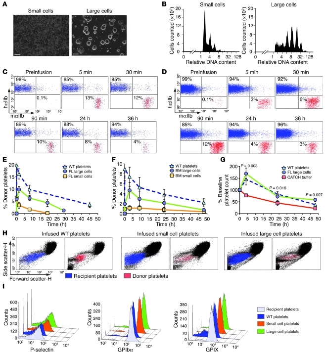
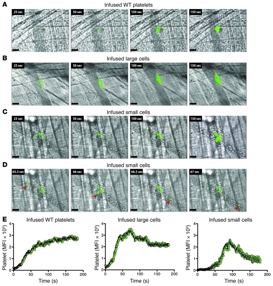
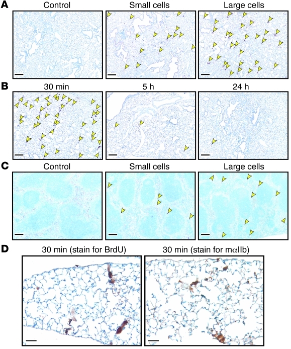
Thrombopoiesis, the process by which circulating platelets arise from megakaryocytes, remains incompletely understood. Prior studies suggest that megakaryocytes shed platelets in the pulmonary vasculature. To better understand thrombopoiesis and to develop a potential platelet transfusion strategy that is not dependent upon donors, of which there remains a shortage, we examined whether megakaryocytes infused into mice shed platelets. Infused megakaryocytes led to clinically relevant increases in platelet numbers. The released platelets were normal in size, displayed appropriate surface markers, and had a near-normal circulating half-life. The functionality of the donor-derived platelets was also demonstrated in vivo. The infused megakaryocytes mostly localized to the pulmonary vasculature, where they appeared to shed platelets. These data suggest that it may be unnecessary to generate platelets from ex vivo grown megakaryocytes to achieve clinically relevant increases in platelet numbers.


Figures

Figure 1
Figure 1. Characterization and infusion of megakaryocytes.
(A) Representative fields of small and large cells. Scale bars: 100 μm. (B) Representative analysis of DNA content of FL small and large cells. (C) Flow cytometry from recipient mouse before and after infusion of 108 WT platelets or (D) 106 FL large cells. (E) Flow cytometric percentage of 106 infused FL large cells and (F) 106 infused adult BM cells. n = 5 for WT platelets, n = 9 for FL cells, n = 5 for BM studies. (G) Percent platelet rise in irradiated thrombocytopenic mice after infusion. n = 5 per arm. Mean ± 1 SD are shown. Initial platelet counts (108/ml) in the 3 groups were: CATCH buffer, 1.8 ± 0.2; platelets, 1.9 ± 0.3; large cells, 1.0 ± 0.2. (H) Size determination of circulating recipient (blue) and infused platelets (red) by forward versus side scatter analysis. (I) Representative flow cytometric analysis of infused and FL-derived platelets comparing P-selectin, GPIbα, and GPIX.
Figure 2
Figure 2. Platelet incorporation into arterial clots after laser injury.
(AC) Representative images of platelets incorporating into clots after infusion of platelets or indicated cells. (A) Donor WT platelets detected using a labeled anti–mouse αIIb Ab. (B and C) Same as in A, but after infusion of FL cells. (D) Sequential stills from left to right noting a recirculating mouse αIIb+ cell (arrowheads) after small cell infusion. Scale bars: 30 μm. (E) Summation of donor platelets incorporated into growing thrombi after infusion of either WT platelets or FL cells. Twenty movies were evaluated per graph.
Figure 3
Figure 3. Organ distribution studies of infused cells.
(A) Staining of lung from hαIIb+ mice infused with saline or small or large cells grown in BrdU (arrows point to stained nuclei). (B) Kinetics of BrdU-labeled large cells in the lungs. (C) Same as in A but for spleen. Scale bars (AC): 200 μm. (D) Large FL cells 30 minutes after infusion in lungs: BrdU-labeled nuclei (left) and mαIIb (right). Scale bars: 50 μm. Data are representative of 3 separate studies.

Comment in

References

    1. Sullivan MT, Cotten R, Read EJ, Wallace EL. Blood collection and transfusion in the United States in 2001. Transfusion. 2007;47(3):385–394. doi: 10.1111/j.1537-2995.2007.01128.x. - DOI - PubMed
    1. Italiano JE, Jr, Lecine P, Shivdasani RA, Hartwig JH. Blood platelets are assembled principally at the ends of proplatelet processes produced by differentiated megakaryocytes. J Cell Biol. 1999;147(6):1299–1312. doi: 10.1083/jcb.147.6.1299. - DOI - PMC - PubMed
    1. Junt T, et al. Dynamic visualization of thrombopoiesis within bone marrow. Science. 2007;317(5845):1767–1770. doi: 10.1126/science.1146304. - DOI - PubMed
    1. Zucker-Franklin D, Philipp CS. Platelet production in the pulmonary capillary bed: new ultrastructural evidence for an old concept. Am J Pathol. 2000;157(1):69–74. - PMC - PubMed
    1. Choi ES, Nichol JL, Hokom MM, Hornkohl AC, Hunt P. Platelets generated in vitro from proplatelet-displaying human megakaryocytes are functional. Blood. 1995;85(2):402–413. - PubMed

Publication types