. 2010 Dec;11(12):1127-35.
doi: 10.1038/ni.1953. Epub 2010 Oct 24.

Critical role for the chemokine receptor CXCR6 in NK cell-mediated antigen-specific memory of haptens and viruses

Affiliations

Critical role for the chemokine receptor CXCR6 in NK cell-mediated antigen-specific memory of haptens and viruses

Silke Paust et al. Nat Immunol. 2010 Dec.

Abstract

Hepatic natural killer (NK) cells mediate antigen-specific contact hypersensitivity (CHS) in mice deficient in T cells and B cells. We report here that hepatic NK cells, but not splenic or naive NK cells, also developed specific memory of vaccines containing antigens from influenza, vesicular stomatitis virus (VSV) or human immunodeficiency virus type 1 (HIV-1). Adoptive transfer of virus-sensitized NK cells into naive recipient mice enhanced the survival of the mice after lethal challenge with the sensitizing virus but not after lethal challenge with a different virus. NK cell memory of haptens and viruses depended on CXCR6, a chemokine receptor on hepatic NK cells that was required for the persistence of memory NK cells but not for antigen recognition. Thus, hepatic NK cells can develop adaptive immunity to structurally diverse antigens, an activity that requires NK cell-expressed CXCR6.


Figures
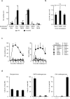
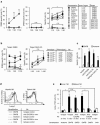
Fig. 1
Fig. 1
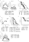
Liver NK-cells develop specific memory to haptens. (a) Hapten-specific CHS responses in naïve Rag2−/−Il2rg−/− recipients of hepatic CD45+NK1.1+Thy1+ NK-cells (1×10⁵) from naïve (vehicle-exposed), DNFB or OXA sensitized Rag1−/− donors. Recipients were challenged 24h or four months post transfer. Hapten-specific ear swelling was determined after 24hrs by subtracting background swelling in naïve mice from that in NK recipients. n=10–15 recipients/group. *p<10−2; **p<10−3 (b) Survival and expansion of adoptively transferred NK-cells is not altered by prior sensitization. Two weeks after the 4 month challenge, recipient mice shown in a were analyzed by FACS and the number of liver-resident CD45+ NK1.1+ cells was determined. Results were similar for DNFB or OXA challenged mice, so data were pooled. No NK1.1+ cells were detected in mock recipient Rag2−/−Il2rg−/− mice (not shown). (c) Liver-restricted memory NK-cells arise in the presence of T and B-cells. Sorted CD45+NK1.1+CD3Thy1+ NK-cells (105) from actin-GFP transgenic donors were transferred to naïve C57BL/6 mice; recipients were challenged six weeks later and analyzed as in a. n=10–15 recipients/group. *p<10−3; **p<10−4 (d) Recruitment of memory NK-cells to sites of challenge is Ag-specific. Hepatic CD45+NK1.1+Thy1+ NK-cells from naïve CD45.1+ WT donors (C57BL/6) and from CD45.2+ DNFB or OXA sensitized WT or actin-GFP transgenic donors were mixed (105 each) and adoptively transferred into naïve Rag2−/−Il2rg−/− recipients. One month post adoptive transfer, recipient ears were challenged with either DNFB or OXA. Livers and ears were harvested at 24, 48 and 72 h, and analyzed for the presence of NK-cells whose origin was distinguished by congenic/fluorescent markers using FACS. No NK-cells were found in acetone challenged control ears (not shown). The mean of all mice analyzed at 24, 48 and 72 h is shown (n=6–7 recipients/group). *p<10−11.
Figure 2
Figure 2
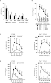
Liver NK-cells develop specific memory to viral Ags. (a) One month after immunization of Rag1−/− mice with viral Ags, 8×10⁴ splenic or hepatic CD45+ NK1.1+ NK-cells were adoptively transferred to naïve Rag2−/−Il2rg−/− mice. Recipient ears were challenged by sq injection two months later. Virus-specific DTH was determined after 24h by subtracting background swelling in naïve mice from that in NK-cell recipients. n=8–10 recipients/group. (b) Influenza-sensitized hepatic NK-cells prolong survival of Rag2−/−Il2rg−/− recipients upon lethal influenza infection. Using the sensitization and transfer protocol in a, recipients were intranasally infected with influenza A PR8 (500pfu) three months after NK-cell transfer. n=15–19 recipients/group. (c) Two month after assessment of DTH, the NK-cell recipients used in a were infected with influenza A PR8 (500pfu), and survival was determined and correlated to DTH. n=19. *p=0.0001 (d) Rag1−/− mice develop virus-specific immunity to influenza and VSV. One month after immunization with influenza VLPs or UV-VSV, Rag1−/− mice were challenged with live virus (2,500 pfu PR8 i.n. or 500 pfu VSV i.v.) and survival was monitored. n=8–12 mice/group. (e) Rag2−/− mice develop long-term protective, virus-specific immunity to VSV. One month after immunization with influenza VLPs or UV-VSV, Rag1−/− mice were challenged by i.m. injection of VSV at LD50 (250pfu) and monitored for survival. n=15–22 mice/group. *p=0.0116 (f) NK-cell memory to influenza A does not require HA. Rag1−/− mice were immunized with HA-containing (PR8) or HA-free (M1) VLPs, challenged one month later and analyzed as in a. n=10–15 mice/group.
Figure 3
Figure 3
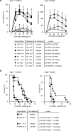
Mouse liver NK-cells recognize and discriminate between HIV-1 and influenza A. (a) One month after subcutaneous immunization of Rag1−/− donor mice with influenza or HIV-1 VLPs, hepatic (left) or splenic (right) CD45+ NK1.1+ NK-cells were adoptively transferred to naïve Rag2−/−Il2rg−/− recipients (8×10⁴ cells/mouse). Two months later, recipients were challenged by subcutaneous VLP injection into one ear. n=12–15 recipients/group. *p<10−2; **p<10−3. (b) NK-cell recognition of HIV-1 derived gag/env containing VLPs and influenza PR8 derived HA and/or M1 containing VLPs occurs independent of genetic background. C57BL/6 Rag1−/− (left) and BALB/c Rag2−/− mice (right) were immunized with VLPs and challenged one month later. In all experiments, background ear swelling in non-immunized mice was determined in parallel and subtracted from measurements in experimental groups. n=10–15 mice/group.
Figure 4
Figure 4
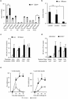
NK cell-expressed CXCR6 is required for NK cell-mediated adaptive immunity to haptens. (a) The percentage of CXCR6-expressing CD45+ NK1.1+ NK-cells from Cxcr6+/− mice on Rag1-sufficient (C57BL/6) and Rag1−/− background were analyzed in different tissues by FACS. LN, lymph node; BM, bone marrow. *p<10−2; **p<10−3; ***p<10−4 (b) 105 NK-cells from DNFB sensitized Rag1−/− Cxcr6+/− donor spleens or livers were sorted for NK1.1 and GFP expression and transferred to naïve Rag2−/−Il2rg−/− recipients. Animals were challenged with DNFB on one ear, and solvent on the other one month later and ear swelling was determined. n=10–12 recipients/group. *p<10−2; **p<10−3; ***p<10−4 (c) Effect of CXCR6 deficiency on DNFB-induced CHS in lympho-competent (C57BL/6, left panel) and Rag1−/− mice (right panel). n=10–12mice/group. (d) Effect of anti-CXCR6 mAb (100 μg/mouse administered i.v. 24h prior to challenge) on DNFB-induced CHS in C57BL/6 (left panel) and Rag1−/− mice (right panel). Hapten sensitized animals were challenged five days post sensitization. n=10–15 mice/group. *p<10−2; **p<10−3; ***p<10−4.
Figure 5
Figure 5
NK cell-expressed CXCR6 is required for NK cell-mediated adaptive immunity to viruses. (a) Effect of anti-CXCR6 mAb (100 μg/mouse 24h prior to challenge) on anti-viral DTH responses in Rag deficient mice on C57BL/6 (left panel) or Balb/c background (right panel). (b) NK cell-mediated protection against lethal influenza A infection depends on CXCR6. Rag−/− mice were immunized and infected one month later with Influenza A PR8 (2,500 pfu for Rag1−/− (left panel) and 10,000 pfu for Rag2−/− (right panel)). Anti-CXCR6 or isotype control mAb (100 μg) was injected on days 1 and 5. n = 8–12 mice/group.
Figure 6
Figure 6
CXCR6 regulates hepatic NK-cell homeostasis. (a) Frequencies of GFP+ and GFP NK-cell subsets were determined in different organs of Cxcr6+/− and Cxcr6−/− mice. NK-cells were identified as CD45+NK1.1+ cells. LN, lymph node; BM, bone marrow. *, p<10−2 **; p<10−3 (b) NK-cell subset ratios in liver and spleen of WT (+/+; C57BL/6), Cxcr6+/− and Cxcr6−/− mice. n=12–15 mice/group. *, p=2×10⁻⁵. (c) Differential distribution of NK-cells recovered from liver or spleen one month after adoptive transfer of sorted subsets (1×10⁵) to Rag2−/−Il2rg−/− recipients. n=10–15 mice/group. *, p<10−3; **, p<10−4 (d) GFP+ but not GFPCxcr6+/− NK-cells outcompete their Cxcr6−/− counterparts upon adoptive transfer. 1×10⁵ GFP+ or GFP CD45+ NK1.1+ NK-cells were sorted from Cxcr6+/− or Cxcr6−/− donors, mixed, transferred to Rag2−/−Il2rg−/− recipients and counted by FACS in liver and spleen two weeks later. n=8–10 recipients/group. *, p<10−3; **, p<10−4 (e) Rag2−/−Il2rg−/− mice received 8×10⁴ DNFB primed CD45+ NK1.1+ GFP+ NK-cells from Cxcr6+/− or Cxcr6−/− donors and were challenged 24h or 1 month later (n=8 mice/group). * p<10−2; **, p<10−3.
Figure 7
Figure 7
Hepatic memory NK-cells mediate hapten-specific killing in vitro. (a) Naive and hapten-sensitized CD45+ NK1.1+ NK-cells were cocultured for 12h with a mixture of two populations of B-cells that were differentially labeled with high or low amounts of CFSE at indicated target to effector (T:E) ratios. CFSElo B-cells served as control, whereas CFSEhi B-cells were either from WT donors and haptenated with DNBS (left and middle panels) or from MHC-I−/− donors (right panel). Hapten-specific killing was determined from the ratio of CFSElo:CFSEhi cells corrected for input. n=10–20 donor mice/group. *, p<10−8; **, p<10−12 (b) Killing capacity of DNFB-primed hepatic CD45+ NK1.1+ NK-cells from Cxcr6+/− or Cxcr6−/− donor mice was determined as in a in the presence of anti-CXCR6 or idotype control mAb. n=12 donor mice/group. *, p<10−2; **, p<10−3; ***, p<10−5. (c) Killing capacity of DNFB primed hepatic CD45+ NK1.1+ NK-cells from Rag1−/− donors at 1:25 T:E ratio was determined in the presence of 10μg/ml anti-CXCR6 or anti-CXCL16 mAb or 500ng/ml CXCL16 and compared to cultures treated with 10μg/ml isotype control. n=15 donor mice/group. *, p<10−2; **, p<10−3. (d,e) NK1.1+ cells were analyzed for binding of anti-Lamp-1 by FACS. Rag−/− donor mice were sensitized with acetone, DNFB or OXA on days 0 and 1, and NK1.1+ NK-cells were sorted from livers or spleens. Some donor mice were injected with 100μg anti-CXCR6 or control mAb 12hrs prior to NK-cell isolation. NK-cells were cocultured with DNBS labeled B-cells in the presence of 10μg/ml FITC-conjugated anti-Lamp-1 with and without addition of 10μg/ml anti-CXCR6 or isotype control. (d) NK-cells were FACS analyzed for anti-Lamp-1 incorporation after three hours. (e) Bars represent means and SEM of pooled data from 3–5 independent experiments; 10–18 donor mice total; 12–20 individual wells per group. *, p<10−9; **.

Comment in

References

    1. Bassing CH, Swat W, Alt FW. The mechanism and regulation of chromosomal V(D)J recombination. Cell. 2002;109(Suppl):S45–S55. - PubMed
    1. Crowle AJ. Delayed hypersensitivity in mice; its detection by skin tests and its passive transfer. Science. 1959;130:159–160. - PubMed
    1. Orme IM, Cooper AM. Cytokine/chemokine cascades in immunity to tuberculosis. Immunol Today. 1999;20:307–312. - PubMed
    1. Dhabhar FS, Satoskar AR, Bluethmann H, David JR, McEwen BS. Stress-induced enhancement of skin immune function: A role for gamma interferon. Proc. Natl. Acad. Sci. U. S. A. 2000;97:2846–2851. - PMC - PubMed
    1. Phanuphak P, Moorhead JW, Claman HN. Tolerance and contact sensitivity to DNFB in mice. II. Specific in vitro stimulation with a hapten, 2,4-dinitrobenzene sulfonic acid (DNBSO3Na) J Immunol. 1974;112:849–851. - PubMed

Publication types

MeSH terms