
The TLX1 oncogene drives aneuploidy in T cell transformation

Kim De Keersmaecker et al. Nat Med. 2010 Nov.

Abstract

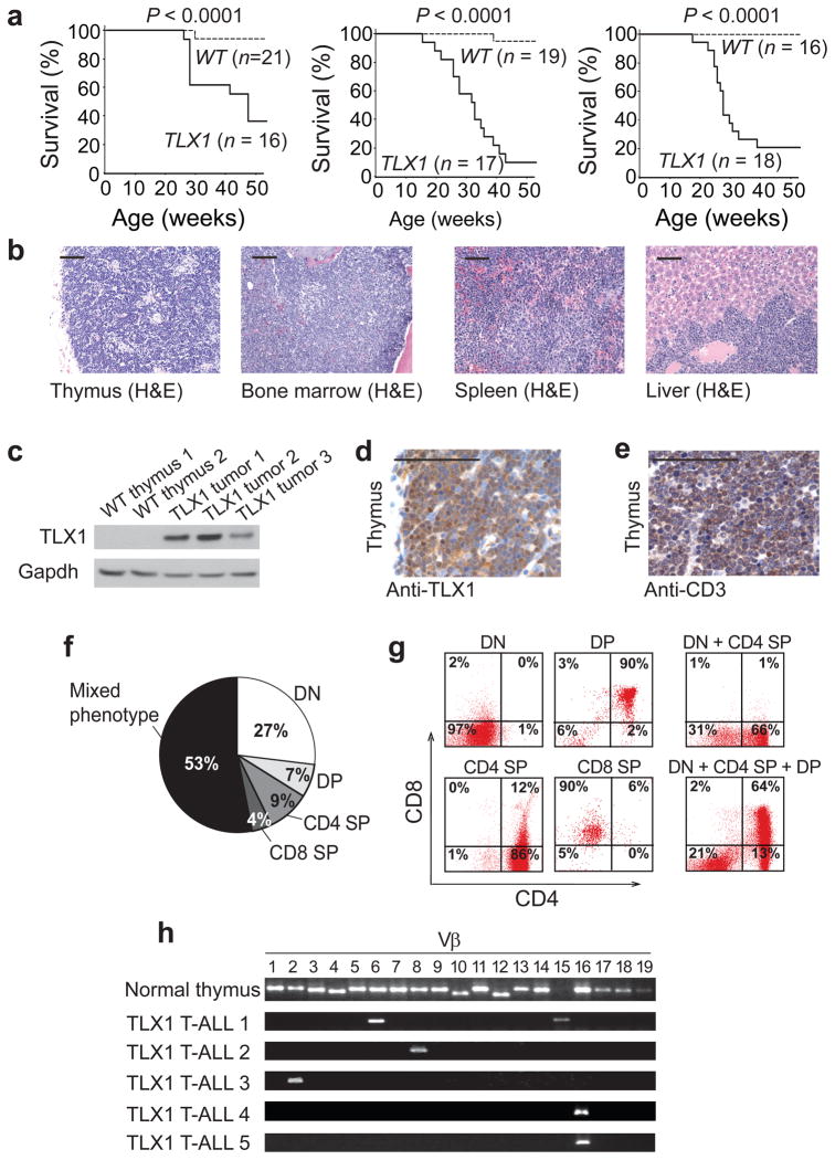
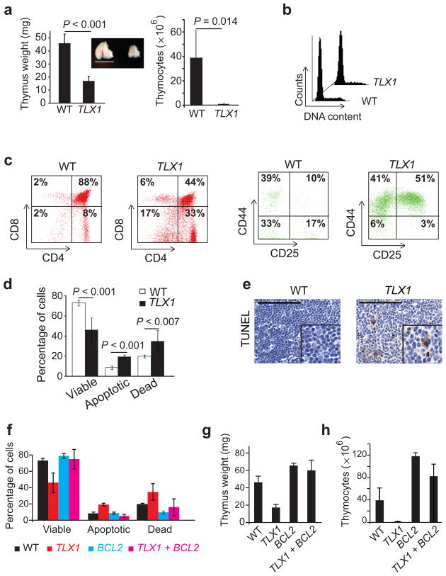
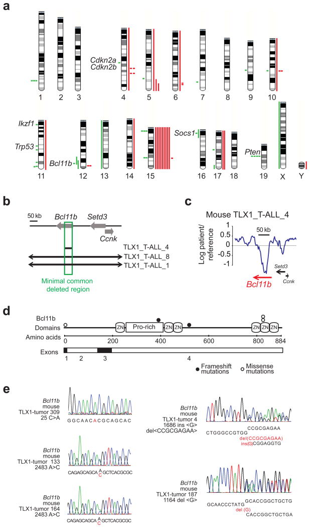
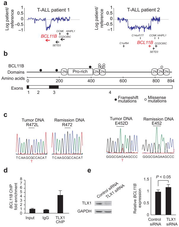
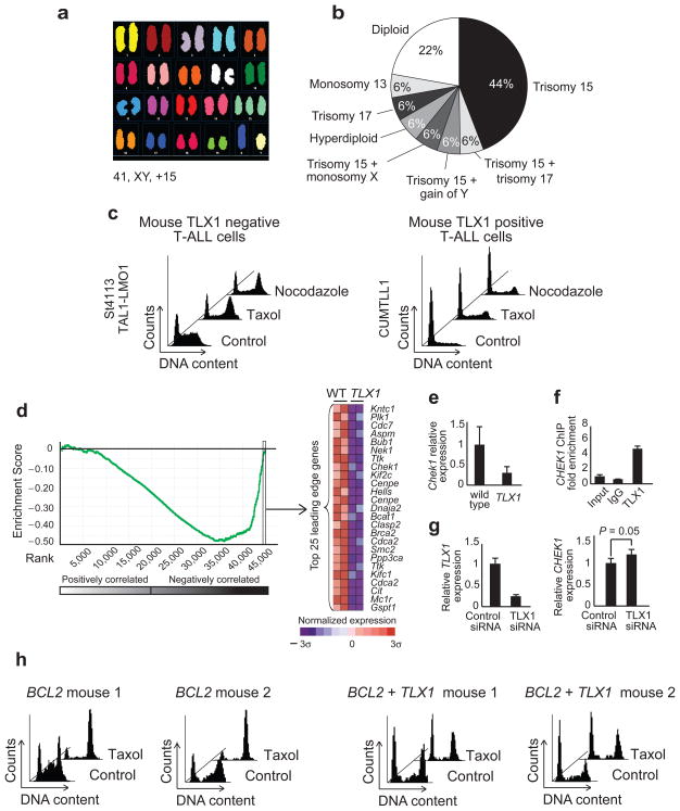
The TLX1 oncogene (encoding the transcription factor T cell leukemia homeobox protein-1) has a major role in the pathogenesis of T cell acute lymphoblastic leukemia (T-ALL). However, the specific mechanisms of T cell transformation downstream of TLX1 remain to be elucidated. Here we show that transgenic expression of human TLX1 in mice induces T-ALL with frequent deletions and mutations in Bcl11b (encoding B cell leukemia/lymphoma-11B) and identify the presence of recurrent mutations and deletions in BCL11B in 16% of human T-ALLs. Most notably, mouse TLX1 tumors were typically aneuploid and showed a marked defect in the activation of the mitotic checkpoint. Mechanistically, TLX1 directly downregulates the expression of CHEK1 (encoding CHK1 checkpoint homolog) and additional mitotic control genes and induces loss of the mitotic checkpoint in nontransformed preleukemic thymocytes. These results identify a previously unrecognized mechanism contributing to chromosomal missegregation and aneuploidy active at the earliest stages of tumor development in the pathogenesis of cancer.


Figures

Fig. 1
Fig. 1
TLX1-induced T-cell leukemias in mice. (a) Kaplan-Meier survival curves of p56Lck-TLX1 transgenic mice and littermate controls from three independent founder lines. Accelerated mortality in TLX1 transgenic mice was associated with the development of immature T-cell tumors. (b) Infiltration of thymus, spleen, bone marrow and liver by immature lymphoblasts. (c) Western blot analysis of TLX1 expression in mouse T-cell tumors. (d-e) Immunohistochemical analysis of TLX1 (d) and CD3 expression in mouse TLX1-induced T-ALL cells (e). Scale bar 100 μm. (f,g) Immunophenotype distribution (f) and representative flow cytometry plots (g) showing heterogeneous expression of CD4 and CD8 in TLX1-induced leukemias. DN, double negative; DP, double positive; SP, single positive. (h) Clonality analysis by expression of TCRB chains in TLX1-induced tumors. Polyclonal expression of TCRB in normal thymocytes is shown as control (upper row).
Fig. 2
Fig. 2
Molecular signatures associated with TLX1-induced transformation. (a) Heat map diagram of the 50 top ranking differentially expressed genes by t-test in mouse TLX1-induced leukemias. (b) GSEA analysis of differentially expressed genes associated with TLX1-induced transformation in mice demonstrates enrichment of this signature in human TLX1/TLX3 expressing T-ALLs. Gene set: Human orthologs of mouse TLX1 signature genes. Data set: TLX1/TLX3 positive vs. TLX1/TLX3 negative human leukemias. Enrichment plots (left) and heat map representations of the 50 top ranking genes in the leading edge (right) are shown. Genes in heat maps are shown in rows, each individual sample is shown in one column. The scale bar shows color coded differential expression from the mean in standard deviation units with red indicating higher levels and blue lower levels of expression.
Fig 3
Fig 3. Developmental defects in thymocyte development in TLX1-transgenic mice
(a) Preleukemic TLX1 transgenic mice at 6 weeks of age showed decreased thymus weight and cellularity compared with littermate controls. Scale bar 10 mm. (b) Cell cycle analysis of control and preleukemic TLX1-transgenic thymocytes via PI staining of DNA content analyzed by flow cytometry. (c) Flow cytometry analysis of T-cell development in preleukemic TLX1-transgenic animals. Accumulation of CD44+CD25+ cells shows a differentiation block at the DN2 stage of thymocyte development. (d) Apoptosis analysis of control and TLX1-transgenic preleukemic thymocytes via annexinV/PI staining. (e) TUNEL staining on thymus tissue sections. Scale bars represent 100 μm. (f-h) Transgenic expression of BCL2 inhibits apoptosis (f) and reverses thymic weight (g) and cellularity (h) in preleukemic TLX1 +BCL2 double transgenic mice.
Fig. 4
Fig. 4
Numerical and structural chromosomal alterations in TLX1-induced mouse T-ALLs. (a) Mouse chromosomal ideogram showing the areas of genetic gain and loss identified by aCGH in TLX1 thymic tumors. Red bars represent areas of gain. Green bars represent areas of copy number loss. (b) Schematic representation of the chromosome 12q commonly deleted region encompassing the Bcl11b locus in mouse TLX1-induced tumors. (c) Array CGH plot showing a focal deletion of the Bcl11b gene in a mouse TLX1-induced T-ALL. (d) Schematic representation of Bcl11b mutations identified in mouse TLX1-induced T-ALLs. (e) DNA sequence chromatograms corresponding to Bcl11b mutations identified in mouse TLX1-induced T-ALLs.
Fig. 5
Fig. 5
BCL11B is a TLX1 target gene mutated in human T-ALL. (a) Array CGH plots showing focal deletions in chromosomal band 14q32.2 encompassing the BCL11B locus in human T-ALL. (b) Schematic representation of BCL11B mutations identified in human T-ALL. (c) DNA sequence analysis of BCL11B in diagnostic and remission T-ALL samples. (d) ChIP analysis of TLX1 binding to BCL11B regulatory sequences in ALL-SIL cells. (e) Western blot analysis of TLX1 and RT-PCR analysis of BCL11B expression in ALL-SIL cells electroporated with control or TLX1 siRNAs.
Fig. 6
Fig. 6
Numerical chromosomal alterations and defects in the mitotic checkpoint in TLX-transgenic mice. (a) SKY analysis of a mouse TLX1-induced tumor with trisomy 15. (b) Distribution of numerical chromosomal aberrations found by SKY and aCGH analysis in mouse TLX1-induced leukemias. (c) Cell cycle analysis of vehicle only, taxol and nocodazole treated mouse T-ALLs showing defective activation of the mitotic checkpoint in mouse TLX1-positive leukemia cells. (d) GSEA analysis of mitotic regulators identified as TLX1 direct target genes by ChIP-chip in sorted thymocytes (DN2 cells) from wild type and preleukemic TLX1 transgenic mice. Gene set: TLX1 direct targets in mitotic cell cycle (GO:0000278). Data set: TLX1 transgenic preleukemic cells vs. wild type. The enrichment plot (left) and heat map representation of the top 25 mitotic genes in the rank of transcripts differentially expressed in TLX1-preleukemic cells (right) are shown. The scale bar at the bottom shows color coded differential expression from the mean in standard deviation units with red indicating higher levels and blue lower levels of expression. (e) RT-PCR analysis of Chek1 expression in thymocytes isolated from wild type or TLX1-transgenic mice. (f) ChIP analysis of TLX1 binding to CHEK1 regulatory sequences in ALL-SIL cells. (g) RT-PCR analysis of TLX1 and CHEK1 expression in ALL-SIL cells electroporated with control or TLX1 siRNAs. (h) Cell cycle analysis of vehicle only and taxol treated mouse thymocytes from BCL2 transgenic and TLX1-BCL2 double transgenic mice showing defective activation of the mitotic checkpoint in mouse TLX1-BCL2-expressing preleukemic cells.

Comment in

  • The T-ALL paradox in cancer.
    Hoang T, Hoang T. Hoang T, et al. Nat Med. 2010 Nov;16(11):1185-6. doi: 10.1038/nm1110-1185. Nat Med. 2010. PMID: 21052064 No abstract available.

Similar articles

Cited by

References

    1. Ferrando AA, et al. Prognostic importance of TLX1 (HOX11) oncogene expression in adults with T-cell acute lymphoblastic leukaemia. Lancet. 2004;363:535–536. - PubMed
    1. Ferrando AA, et al. Gene expression signatures define novel oncogenic pathways in T cell acute lymphoblastic leukemia. Cancer Cell. 2002;1:75–87. - PubMed
    1. Kees UR, et al. Expression of HOX11 in childhood T-lineage acute lymphoblastic leukaemia can occur in the absence of cytogenetic aberration at 10q24: a study from the Children's Cancer Group (CCG) Leukemia. 2003;17:887–893. - PubMed
    1. Berger R, et al. t(5;14)/HOX11L2-positive T-cell acute lymphoblastic leukemia. A collaborative study of the Groupe Francais de Cytogenetique Hematologique (GFCH) Leukemia. 2003;17:1851–1857. - PubMed
    1. Bernard OA, et al. A new recurrent and specific cryptic translocation, t(5;14)(q35;q32), is associated with expression of the Hox11L2 gene in T acute lymphoblastic leukemia. Leukemia. 2001;15 :1495–1504. - PubMed

Publication types

MeSH terms