. 2010 Nov;17(11):1312-7.
doi: 10.1038/nsmb.1938. Epub 2010 Oct 24.

Structural and mechanistic insights into cooperative assembly of dimeric Notch transcription complexes

Affiliations

Structural and mechanistic insights into cooperative assembly of dimeric Notch transcription complexes

Kelly L Arnett et al. Nat Struct Mol Biol. 2010 Nov.

Abstract

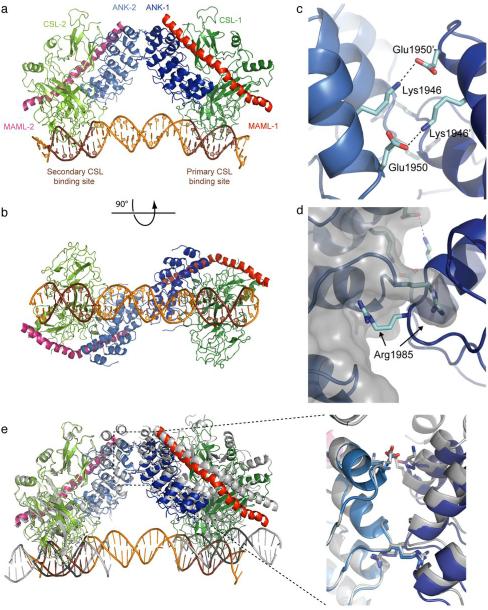
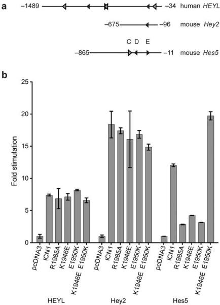
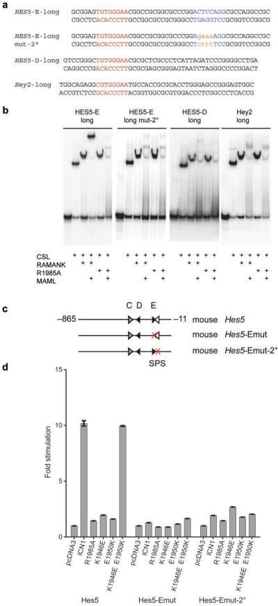
Ligand-induced proteolysis of Notch produces an intracellular effector domain that transduces essential signals by regulating the transcription of target genes. This function relies on the formation of transcriptional activation complexes that include intracellular Notch, a Mastermind co-activator and the transcription factor CSL bound to cognate DNA. These complexes form higher-order assemblies on paired, head-to-head CSL recognition sites. Here we report the X-ray structure of a dimeric human Notch1 transcription complex loaded on the paired site from the human HES1 promoter. The small interface between the Notch ankyrin domains could accommodate DNA bending and untwisting to allow a range of spacer lengths between the two sites. Cooperative dimerization occurred on the human and mouse Hes5 promoters at a sequence that diverged from the CSL-binding consensus at one of the sites. These studies reveal how promoter organizational features control cooperativity and, thus, the responsiveness of different promoters to Notch signaling.


Figures

Figure 1
Figure 1
Structure of a dimer of Notch transcription complexes on paired-site DNA. (a) Ribbon representation of two copies of the Notch1 ANK–CSL–MAML1 complex on the paired-site DNA element from the human HES1 promoter. The NTC complex bound to the primary CSL binding site (TGTGGGAA) contains ANK (blue), CSL (green), and MAML1 (red). The complex bound to the secondary CSL binding site (CGTGTGAA) includes a second molecule of ANK (light blue), CSL (chartreuse) and MAML1 (pink). The DNA is colored orange with the two CSL binding sites highlighted in brown. (b) A view related by a 90° rotation around the DNA axis of panel (a). (c, d) Interactions in the NTC dimer interface, including salt-bridges between Lys 1946 of one Notch ANK domain and Glu 1950 of the other ANK domain (c), and between Arg 1985 of one Notch ANK domain and a pocket created by Arg 1985 and surrounding residues in the other ANK domain (d). (e) Comparison of the NTC dimeric structure to the crystallographic pseudo-dimer seen for two symmetry mates in 2F8X (grey). The panel to the right shows a zoomed view of the superimposed dimer interface.
Figure 2
Figure 2
NTC dimer formation on putative Notch targets hHES4 and hFJX1 and on SPS elements with 15–17 bp spacers. (a) Oligonucleotide duplexes used for Electrophoretic Mobility Shift Assays. (b) EMSAs performed using CSL, MAML1 and wild-type Notch1 RAMANK or R1985A on SPS elements from the promoters of putative Notch target genes, hHES4 and hFJX1. Schematic representation of the complexes assembled include CSL (green), RAMANK (blue) and MAML1 (red) on DNA (grey). (c) EMSAs performed on DNA with CSL sites in the SPS arrangement with spacers of 16 bp (the HES1 SPS), 15 bp (from the SYT14 promoter) and 17 bp (from the CUL1 promoter).
Figure 3
Figure 3
Dimer-dependent and dimer-independent DNA elements identified in the promoters of Notch responsive genes hHEYL, mHey2 and mHes5. (a) Diagram of the promoter elements used to control the expression of luciferase reporter genes. Predicted high affinity CSL binding sites (black arrowheads) and low affinity sites (grey arrowheads) are depicted with the direction of the arrowhead representing the relative orientation of the CSL binding site. (b) Responsiveness of luciferase reporter genes under control of hHEYL, mHey2, or mHes5 promoter elements upon expression of various activated Notch1 receptors (see methods). Fold stimulation is expressed relative to empty vector control, which is set to a value of 1. Error bars correspond to standard deviations.
Figure 4
Figure 4
The mouse and human HES5 proximal promoters each contain two conserved high affinity CSL binding sites, one of which is essential for activation of expression. (a) HES5 D and E site single CSL site oligonucleotide duplexes (which have identical CSL binding sites and are conserved between mouse and human) used for EMSAs. (b) EMSAs performed on single CSL site DNA from the D and E sites of the human HES5 promoter. (c) Diagram of the promoter elements used to control expression in luciferase reporter constructs. Predicted high affinity CSL binding sites (black arrowheads) and low affinity sites (grey arrowheads) are depicted with the direction of the arrowhead representing the relative orientation of the CSL binding site. Mutated CSL binding sites are indicated by a red X. (d) Luciferase reporter assays performed on the Hes5-Dmut and Hes5-Emut response elements using the indicated forms of ICN1. Fold stimulation is expressed relative to empty pcDNA3 vector control, which is set to a value of 1. Error bars represent standard deviations.
Figure 5
Figure 5
Identification of a non-consensus SPS element in the HES5 promoter. (a) Long oligonucleotide duplexes used for EMSAs, spanning the primary CSL binding site and flanking sequence. 2°: secondary site. (b) EMSAs performed on the DNA sequences in (a) using CSL, MAML1 and wild-type Notch1 RAMANK or R1985A RAMANK as indicated. (c) Diagram of the promoter elements used to control expression in luciferase reporter constructs. Predicted high affinity CSL binding sites (black arrowheads), low affinity sites (grey arrowheads) and the cryptic binding site (white arrowhead) are depicted with the direction of the arrowhead representing the relative orientation of the CSL binding site. Mutated CSL binding sites are indicated by a red X. (d) Luciferase reporter assays, performed on the normal mHes5 promoter or on promoters with mutations at the primary or cryptic secondary CSL binding sites (mHes5-Emut or mHes5-Emut-2°) using the indicated forms of ICN1. Fold stimulation is expressed relative to empty pcDNA3 vector control, which is set to a value of 1. Error bars represent standard deviations.

Similar articles

Cited by

References

    1. Gridley T. Notch signaling and inherited disease syndromes. Hum Mol Genet. 2003;12(Spec No 1):R9–13. - PubMed
    1. Weng AP, et al. Activating mutations of NOTCH1 in human T cell acute lymphoblastic leukemia. Science. 2004;306:269–71. - PubMed
    1. Brown MS, Ye J, Rawson RB, Goldstein JL. Regulated intramembrane proteolysis: a control mechanism conserved from bacteria to humans. Cell. 2000;100:391–8. - PubMed
    1. Mumm JS, et al. A ligand-induced extracellular cleavage regulates gamma-secretase-like proteolytic activation of Notch1. Mol Cell. 2000;5:197–206. - PubMed
    1. Brou C, et al. A novel proteolytic cleavage involved in Notch signaling: the role of the disintegrin-metalloprotease TACE. Mol Cell. 2000;5:207–16. - PubMed

Publication types

MeSH terms

Associated data