. 2010 Nov;17(11):1292-7.
doi: 10.1038/nsmb.1917. Epub 2010 Oct 24.

Molecular organization of the COG vesicle tethering complex

Affiliations

Molecular organization of the COG vesicle tethering complex

Joshua A Lees et al. Nat Struct Mol Biol. 2010 Nov.

Abstract

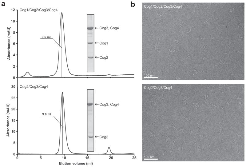
Multisubunit tethering complexes of the CATCHR (complexes associated with tethering containing helical rods) family are proposed to mediate the initial contact between an intracellular trafficking vesicle and its membrane target. To begin elucidating the molecular architecture of one well-studied example, the conserved oligomeric Golgi (COG) complex, we reconstituted its essential subunits (Cog1, Cog2, Cog3 and Cog4) and used single-particle electron microscopy to reveal a y-shaped structure with three flexible, highly extended legs. Labeling experiments established that the N termini of all four subunits interact along the proximal segment of one leg, whereas three of the four C termini are located at the tips of the legs. Our results suggest that the central region of the Cog1-Cog2-Cog3-Cog4 complex, as well as the distal regions of at least two legs, all participate in interactions with other components of the intracellular trafficking machinery.


Figures

Figure 1
Figure 1
Analysis of the quadruple knockout strain for growth and trafficking defects. (a) Δcog5 Δcog6 Δcog7 Δcog8 cells displayed no growth defect or temperature sensitivity. Each growth curve represents either the haploid wild-type (open symbols) or Δcog5 Δcog6 Δcog7 Δcog8 (filled symbols) strain. Error bars represent the standard deviation (s.d.) for three independent cultures. (b) Haploid strains were grown on solid GNA medium at the indicated temperatures and exposed to nitrocellulose membranes; membranes were immunoblotted for secreted Kar2. erd2-B36 and erd2-B25 strains contain mutations in the HDEL receptor, responsible for the retrieval of ER-localized proteins, and therefore display high levels of inappropriate Kar2 secretion.
Figure 2
Figure 2
Purification and electron microscopy of the COG subcomplexes. (a) Gel filtration of purified Cog2-4 and Cog1-4 subcomplexes. Purified samples were analyzed on a Superdex 200 10/300 GL column; elution volume is indicated for each complex. Insets: Coomassie-stained gels for each purified complex. Note that Cog3 and Cog4 co-migrate as a single band. (b) EM analysis of purified Cog2-4 and Cog1-4. Representative raw images of negatively stained particles are shown. The scale bars represent 100 nm.
Figure 3
Figure 3
Projection structures of the Cog2-4 and Cog1-4 (“core”) complexes. (a) Selected class averages from purified Cog1-4 and Cog2-4 complexes. The number in the bottom right-hand corner of each panel indicates the number of particles in the class average. The side length of each panel is approximately 45 nm. (b) Cartoon representation of Cog1-4 denoting key structural elements. (c) Length measurements of Cog1-4 leg structures. Bar graph represents length measurements from raw particles using ImageJ (± s.d.; n=3). Lengths are also represented in b. (d) The thicknesses of the legs of the complex in class averages were measured and compared using ImageJ at the points indicated on the cartoon. Notable is the increased thickness of leg A (position 1) relative to the others and the presence of a swelling on leg B (position 3) (± s.d.; n=12). (e) The x-ray structure of the CATCHR-family subunit Exo70, provided for comparison as described in the text.
Figure 4
Figure 4
Model for subunit organization in the Cog1-4 core complex. (a) Localization of GFP tags from GFP-labeled Cog1-4 complexes. Four representative class averages are shown for each tagged complex. The tag position is indicated by an arrowhead in one of the four averages. The Cog4 C-terminus was localized by using a C-terminally truncated version of Cog4 (residues 1–553) carrying an N-terminal GFP tag. The inferred C-terminus of Cog4 is indicated by a white arrowhead. The number in the bottom right-hand corner of each panel indicates the number of particles in the class average. The side length of each panel is approximately 45 nm. (b) Model for subunit localization in the Cog1-4 complex; arrowheads indicate C-termini.
Figure 5
Figure 5
Functional architecture of the Cog1-4 core complex. (a) Selected class averages of the Cog1/2/3/4/8 complex are shown (side length = 50 nm); the Cog4 N-terminus carries a GFP tag. Cog8 forms a curved extension from the tip of the Cog1 foot structure and ends in a globular structure. Also shown is a class average from the Cog1-GFP/2/3/4 complex, in which a pronounced gap between the tip of the Cog1 foot and the C-terminal GFP tag is evident. The number in the bottom right-hand corner of each panel indicates the number of particles in the class average. (b) Cartoon representation of the Cog1-4 core complex colored by subunit, with expanded views highlighting functionally important regions. The crystal structure of human Cog4 (C-terminal residues 537–785) is shown, together with a magnified image of the salt bridge interaction between Arg 729 and Glu 764 that positions and/or stabilizes the C-terminal domain. The N-terminal subunit interaction region is also highlighted, together with the approximate inferred interaction sites for human Syntaxin 5 and Sly1,. Finally, the NMR structure of yeast Cog2 (C-terminal residues 109–262) is shown, indicating its approximate position within the core complex.

References

    1. Whyte JR, Munro S. Vesicle tethering complexes in membrane traffic. J Cell Sci. 2002;115:2627–2637. - PubMed
    1. Cai H, Reinisch K, Ferro-Novick S. Coats, tethers, Rabs, and SNAREs work together to mediate the intracellular destination of a transport vesicle. Dev Cell. 2007;12:671–682. - PubMed
    1. Sztul E, Lupashin V. Role of vesicle tethering factors in the ER-Golgi membrane traffic. FEBS Lett. 2009;583:3770–83. - PMC - PubMed
    1. Yu I, Hughson FM. Tethering factors as organizers of intracellular vesicular traffic. Ann Rev Cell Dev Biol. 2010;26 in press. - PubMed
    1. Ungar D, Oka T, Krieger M, Hughson FM. Retrograde transport on the COG railway. Trends Cell Biol. 2006;16:113–120. - PubMed

Publication types

MeSH terms

Substances