. 2011 Jul;68(14):2453-67.
doi: 10.1007/s00018-010-0564-7. Epub 2010 Oct 24.

The cross-talk between the urokinase receptor and fMLP receptors regulates the activity of the CXCR4 chemokine receptor

Affiliations

The cross-talk between the urokinase receptor and fMLP receptors regulates the activity of the CXCR4 chemokine receptor

Nunzia Montuori et al. Cell Mol Life Sci. 2011 Jul.

Abstract

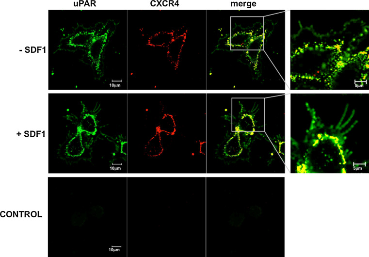
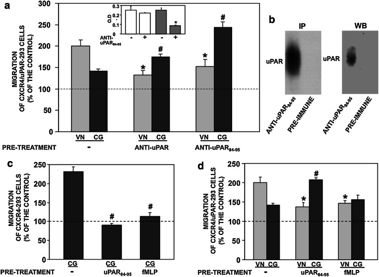
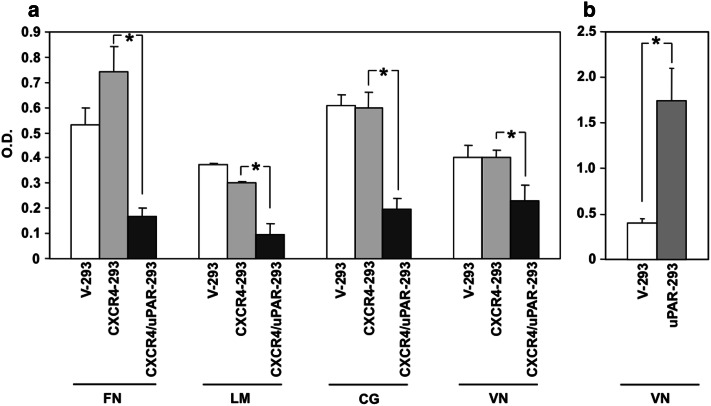
The receptor (CXCR4) for the stromal-derived factor-1 (SDF1) and the urokinase-receptor (uPAR) are up-regulated in various tumors. We show that CXCR4-transfected cells migrate toward SDF1 on collagen (CG) and do not on vitronectin (VN). Co-expression of cell-surface uPAR, which is a VN receptor, impairs SDF1-induced migration on CG and allows migration on VN. Blocking fMLP receptors (fMLP-R), alpha-v integrins or the uPAR region capable to interact with fMLP-Rs, impairs migration of uPAR/CXCR4-transfected cells on VN and restores their migration on CG. uPAR co-expression also reduces the adherence of CXCR4-expressing cells to various components of the extracellular matrix (ECM) and influences the partitioning of beta1 and alpha-v integrins to membrane lipid-rafts, affecting ECM-dependent signaling. uPAR interference in CXCR4 activity has been confirmed in cells from prostate carcinoma. Our results demonstrate that uPAR expression regulates the adhesive and migratory ability of CXCR4-expressing cells through a mechanism involving fMLP receptors and alpha-v integrins.


Figures

Fig. 1
Fig. 1
uPAR and CXCR4 are expressed by transfected HEK-293 cells. a HEK-293 cells were transfected with CXCR4 cDNA (CXCR4-293), with uPAR and CXCR4 cDNAs (CXCR4/uPAR-293), or with empty vectors (V-293). Transfected cells were analyzed by flow cytometry with anti-CXCR4 antibodies, anti-uPAR antibodies or nonimmune immunoglobulins (Ig). b V-293, CXCR4-293 and CXCR4/uPAR-293 cells were lysed in 1% TRITON X-100 and 5 μg of proteins were analyzed by Western blot with uPAR-specific antibodies. uPAR, expressed only in CXCR4/uPAR-293 cells, was mainly in the full-length form
Fig. 2
Fig. 2
uPAR and CXCR4 colocalize on the surface of transfected HEK-293 cells. HEK-293 cells transfected with CXCR4 and uPAR cDNAs (CXCR4/uPAR-293) were grown on glass slides and stimulated with SDF1 or buffer for 1 h. Then cells were incubated with the mouse uPAR-specific antibody (green) and the rabbit CXCR4-specific antibody (red), and analyzed by confocal microscopy. SDF1-treated cells, incubated with nonimmune Ig and secondary antibodies, were shown as control. Scale bar 10 μm
Fig. 3
Fig. 3
uPAR expression affects SDF1-induced cell migration on VN and CG. a HEK-293 cells transfected with CXCR4 cDNA (CXCR4-293), uPAR and CXCR4 cDNAs, (CXCR4/uPAR-293) or empty vectors (V-293), were plated in Boyden chambers on filters coated with fibronectin (FN), laminin (LM), collagen (CG) or vitronectin (VN), and allowed to migrate in the absence of chemoattractants. The values are the mean ± SD of three experiments performed in triplicate. b Transfected cells were plated in Boyden chambers and allowed to migrate toward 100 ng/ml SDF1 on filters coated with FN, LM, CG, or VN. 100% values represent cell migration in the absence of chemoattractants. The values are the mean ± SD of three experiments performed in triplicate. *p ≤ 0.05 as determined by Student’s t test. c CXCR4/uPAR-293 cells were plated in Boyden chambers and allowed to migrate toward 100 ng/ml SDF1 on filters coated with CG or VN, in the presence or in the absence of 10 nM of the aminoterminal fragment of uPA (ATF). ATF was added to both upper and lower compartments of the Boyden chamber. 100% values represent cell migration in the absence of chemoattractants. The values are the mean ± SD of three experiments. *p ≤ 0.05 as determined by Student’s t test
Fig. 4
Fig. 4
uPAR effects on SDF1-induced migration involve the uPAR84–95 epitope and fMLP-receptors. a CXCR4/uPAR-293 cells were incubated with nonimmune immunoglobulins (−), anti-uPAR antibodies or anti-uPAR84–95 antibodies, plated in Boyden chambers and allowed to migrate toward 100 ng/ml SDF1 on filters coated with collagen (CG) and vitronectin (VN). 100% values represent cell migration in the absence of chemoattractants. The values are the mean ± SD of three experiments performed in triplicate. *p ≤ 0.05, as determined by Student’s t test, compared to migration on VN of cells treated with nonimmune Ig. # p ≤ 0.05, as determined by Student’s t test, compared to migration on CG of cells treated with nonimmune Ig. Inset CXCR4/uPAR-293 cells were plated on VN-coated wells in the presence of 5 μg/ml of nonimmune immunoglobulins (−) or the anti-uPAR84–95 polyclonal antibody (white columns) or of 50 μg/ml of nonimmune immunoglobulins (−) or the anti-uPAR84–95 polyclonal antibody (grey columns). The attached cells were fixed and stained with crystal violet. The stain was eluted, and the absorbance at 540 nm was measured with a spectrophotometer. The values represent the means ± SD of three experiments performed in triplicate. *p ≤ 0.05, as determined by Student’s t test. b Characterization of the anti-uPAR84–95 polyclonal antibody: cell-surface proteins of uPAR-transfected 293 cells were biotinylated; cells were lysed and 500 μg of proteins were immunoprecipitated (IP) with 10 μg/ml of the anti-uPAR84–95 polyclonal antibody or pre-immune immunoglobulins (left). uPAR-293 cells were lysed in TRITON X-100 and 5 μg of proteins were analyzed by Western blot (WB) with 10 μg/ml of the anti-uPAR84–95 polyclonal antibody (right). c CXCR4-293 cells were pre-incubated with buffer (−), 100 nM uPAR84–95 peptide or 100 nM fMLP, plated in Boyden chambers and allowed to migrate toward 100 ng/ml SDF1 on CG-coated filters. 100% values represent cell migration in the absence of chemoattractants. The values are the mean ± SD of three experiments performed in triplicate. # p ≤ 0.05, as determined by Student’s t test, compared to migration on CG of untreated cells. d CXCR4/uPAR-293 cells were pre-incubated with buffer (−), 100 nM uPAR84–95 peptide or 100 nM fMLP, plated in Boyden chambers and allowed to migrate toward 100 ng/ml SDF1 on filters coated with CG or VN. 100% values represent cell migration in the absence of chemoattractants. The values are the mean ± SD of three experiments performed in triplicate. *p ≤ 0.05, as determined by Student’s t test, compared to migration on VN of untreated cells. # p ≤ 0.05, as determined by Student’s t test, compared to migration on CG of untreated cells
Fig. 5
Fig. 5
uPAR84–95 peptide and fMLP prevent agonist-FPR interaction. All fMLP-receptors expressed by HEK-293 are involved in uPAR effects on SDF1-induced migration. a Representative confocal images of CXCR4/uPAR-293 cells incubated with buffer (none), 100 nM fMLP, 100 nM uPAR84–95 peptide or 100 nM scrambled peptide for 30 min at 37°C and exposed for further 30 min at 37°C to 10 nM N-formyl-Nle-Leu-Phe-Nle-Tyr-Lys-fluorescein. Z-series images represent focal planes corresponding to 0.5 μm vertical interval. Original magnification 630×. Scale bar 10 μm. b CXCR4/uPAR-293 cells were pre-incubated with buffer (−) or 5 nM WKYMVm peptide, plated in Boyden chambers and allowed to migrate toward 100 ng/ml SDF1 on filters coated with collagen (CG) or vitronectin (VN). 100% values represent cell migration in the absence of chemoattractants. The values are the mean ± SD of three experiments. *p ≤ 0.05, as determined by Student’s t test. Inset 5 μg of total RNA from CXCR4/uPAR-293 cells was reversely transcribed and amplified using FPRL2-specific primers. The reaction products were analyzed by electrophoresis and stained with ethidium bromide, followed by photography under ultraviolet illumination
Fig. 6
Fig. 6
uPAR effects on SDF1-induced migration involve specific integrins. CXCR4/uPAR-293 cells were incubated with nonimmune Ig (−) or an anti-alpha-v monoclonal antibody, plated in Boyden chambers and allowed to migrate toward 100 ng/ml SDF1 on filters coated with collagen (CG) or vitronectin (VN). 100% values represent cell migration in the absence of chemoattractants. The values are the mean ± SD of three experiments performed in triplicate. *p ≤ 0.05, as determined by Student’s t test
Fig. 7
Fig. 7
uPAR expression down-regulates the adhesion of CXCR4-transfected cells to ECM components. a CXCR4-293, CXCR4/uPAR-293 or control cells (V-293) were plated on FN-, LM-, CG-, or VN-coated wells. The attached cells were fixed and stained with crystal violet. The stain was eluted, and the absorbance at 540 nm was measured by a spectrophotometer. The values represent the mean ± SD of six experiments performed in triplicate. *p ≤ 0.05, as determined by Student’s t test. b HEK-293 cells transfected with uPAR cDNA (uPAR-293) or control cells transfected with the empty vector (V-293) were plated on VN-coated wells. The attached cells were fixed and stained with crystal violet. The stain was eluted, and the absorbance at 540 nm was measured by a spectrophotometer. The values represent the mean ± SD of three experiments performed in triplicate. *p ≤ 0.05, as determined by Student’s t test
Fig. 8
Fig. 8
uPAR expression affects integrin association to lipid rafts. CXCR4-293 and CXCR4/uPAR-293 cells were plated on CG- or VN-coated wells, incubated for 1 h at 37°C and lysed in buffer containing 1% Triton X-100. Cell lysates were subjected to sucrose density gradient ultracentrifugation. After centrifugation, 8 ml was harvested as 1-ml fractions. Equal volumes of each fraction were analyzed by Western blot with anti-beta1 or anti-alpha-v integrin antibodies or with anti-CXCR4 antibodies (upper panels) or with anti-uPAR or anti-caveolin antibodies (lower panels)
Fig. 9
Fig. 9
uPAR expression affects ECM signaling in CXCR4 expressing cells. CXCR4-293 (left) and CXCR4/uPAR-293 (right) cells were serum-starved and plated on collagen (CG) (upper panels) or vitronectin (VN) (lower panels). The cells were harvested at the indicated times and lysed for Western-blot analysis with anti-phospho-ERKs and anti-ERK 2 (as a loading control) antibodies
Fig. 10
Fig. 10
uPAR expression regulates SDF1-induced migration in tumor cells. a PC3 cells were pre-incubated with nonimmune Ig (−) or the polyclonal anti-uPAR84–95 antibody, plated in Boyden chambers and allowed to migrate toward 100 ng/ml SDF1 on filters coated with collagen (CG) or vitronectin (VN). b PC3 cells were pre-incubated with buffer (−) or the uPAR84–95 peptide, plated in Boyden chambers and allowed to migrate toward 100 ng/ml SDF1 on filters coated with CG or VN. c PC3 cells were transiently transfected with a uPAR-specific siRNA (+) or a control siRNA (−). Transfected cells were plated in Boyden chambers and allowed to migrate toward 100 ng/ml SDF1 on filters coated with CG or VN (left). 100% values represent cell migration in the absence of chemoattractants. The values are the mean ± SD of three experiments performed in triplicate. *p ≤ 0.05, as determined by Student’s t test. uPAR silencing was assessed by Western-blot analysis of transfected cell lysates with uPAR-specific antibodies (right)

Similar articles

Cited by

References

    1. Murdoch C. CXCR4: chemokine receptor extraordinaire. Immunol Rev. 2000;177:175–184. doi: 10.1034/j.1600-065X.2000.17715.x. - DOI - PubMed
    1. Balkwill F. The significance of cancer cell expression of the chemokine receptor CXCR4. Semin Cancer Biol. 2004;14:171–179. doi: 10.1016/j.semcancer.2003.10.003. - DOI - PubMed
    1. Kucia M, Reca R, Miekus K, Wanzeck J, Wojakowski W, Janowska-Wieczorek A, Ratajczak J, Ratajczak MZ. Trafficking of normal stem cells and metastasis of cancer stem cells involve similar mechanisms: pivotal role of the SDF-1-CXCR4 axis. Stem Cells. 2005;23:879–894. doi: 10.1634/stemcells.2004-0342. - DOI - PubMed
    1. Zlotnik A. Chemokines and cancer. Int J Cancer. 2006;119:2026–2029. doi: 10.1002/ijc.22024. - DOI - PubMed
    1. Burger JA, Kipps TJ. CXCR4: a key receptor in the crosstalk between tumor cells and their microenvironment. Blood. 2006;107:1761–1767. doi: 10.1182/blood-2005-08-3182. - DOI - PubMed

Publication types

MeSH terms

LinkOut - more resources