. 2010 Dec 7;49(48):10287-97.
doi: 10.1021/bi100500m. Epub 2010 Nov 12.

Intrinsically disordered PEP-19 confers unique dynamic properties to apo and calcium calmodulin

Affiliations

Intrinsically disordered PEP-19 confers unique dynamic properties to apo and calcium calmodulin

Xu Wang et al. Biochemistry. .

Abstract

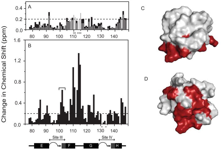
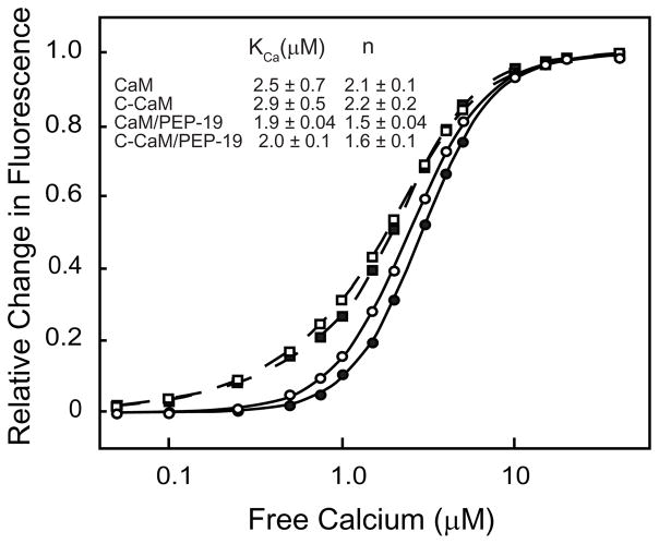
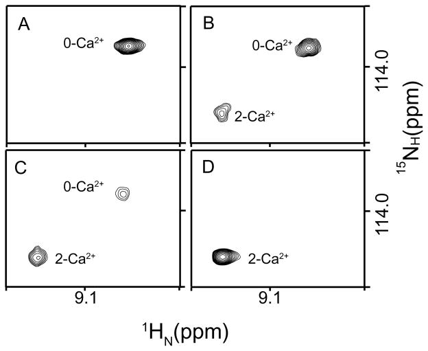
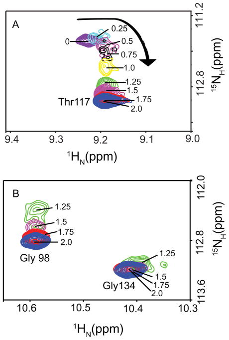
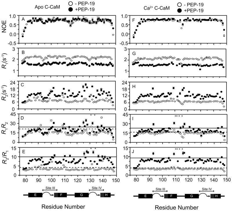
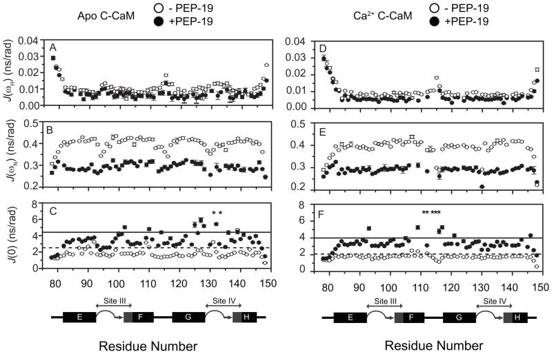
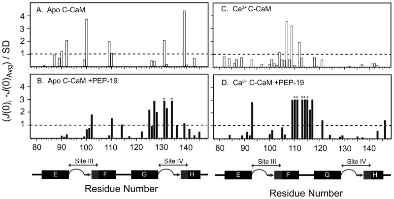
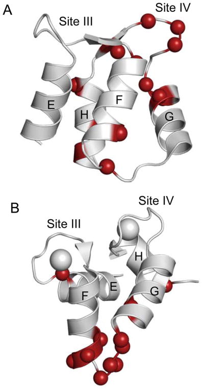
PEP-19 (Purkinje cell protein 4) is an intrinsically disordered protein with an IQ calmodulin (CaM) binding motif. Expression of PEP-19 was recently shown to protect cells from apoptosis and cell death due to Ca(2+) overload. Our initial studies showed that PEP-19 causes novel and dramatic increases in the rates of association of Ca(2+) with and dissociation of Ca(2+) from the C-domain of CaM. The goal of this work was to study interactions between the C-domain of CaM (C-CaM) and PEP-19 by solution nuclear magnetic resonance (NMR) to identify mechanisms by which PEP-19 regulates binding of Ca(2+) to CaM. Our results show that PEP-19 causes a greater structural change in apo C-CaM than in Ca(2+)-C-CaM, and that the first Ca(2+) binds preferentially to site IV in the presence of PEP-19 with exchange characteristics that are consistent with a decrease in Ca(2+) binding cooperativity. Relatively weak binding of PEP-19 has distinct effects on chemical and conformational exchange on the microsecond to millisecond time scale. In apo C-CaM, PEP-19 binding causes a redistribution of residues that experience conformational exchange, leading to an increase in the number of residues around Ca(2+) binding site IV that undergo conformational exchange on the microsecond to millisecond time scale. This appears to be caused by an allosteric effect because these residues are not localized to the PEP-19 binding site. In contrast, PEP-19 increases the number of residues that exhibit conformational exchange in Ca(2+)-C-CaM. These residues are primarily localized to the PEP-19 binding site but also include Asp93 in site III. These results provide working models for the role of protein dynamics in the regulation of binding of Ca(2+) to CaM by PEP-19.


Figures

Figure 1
Figure 1. Effect of PEP-19 on C-CaM amides in the absence and presence of Ca2+
(A) Amide chemical shift perturbations (Δδavg) for Ca2+-bound C-CaM at 298 K are shown by black bars. Grey bars show Δδavg that result from binding PEP-19 to the C-domain of intact Ca2+-CaM at 320 K (14). Asterisks denote resonances with significant line-broadening in the presence of PEP-19. (B) Δδavg for apo C-CaM at 298 K. Panels (C) and (D) map residues with Δδavg >0.2 ppm on the solvent accessible surfaces of Ca2+-CaM (pdb:1J7P) and apo-C-CaM (pdb:1F71), respectively. The structures are presented in the same relative orientation. The diagram below panel B shows the relative positions of the helices E to H in the primary sequence of CaM. The 12 amino acids Ca2+ binding sites III and IV are also shown in grey. Coil, β-strand and α-helical portions of these structures are indicated by loops, arrows, and boxes, respectively.
Figure 2
Figure 2. Effect of PEP-19 on the Ca2+ binding constant (Kca) and Hill coefficient (n)
Intrinsic tyrosine fluorescence was monitored during titration of CaM (○), CaM+PEP-19 (□), C-CaM (●), and C-CaM/PEP-19 (■) with Ca2+. The lines show fits of the data to the Hill equation (Eq.1).
Figure 3
Figure 3. Ca2+ -dependent changes in amide chemical shifts of C-CaM show strong cooperativity
Resonance of Thr117 in the linker region of C-CaM exhibited two-state slow exchange at various molar ratio of Ca2+ added into C-CaM, indicating highly cooperative Ca2+ binding to sites III and IV. (A) [Ca2+]/[C-CaM]=0; (B) [Ca2+]/[C-CaM]=0.3; (C) [Ca2+]/[C-CaM]=0.6; (D) [Ca2+]/[C-CaM]=2.0.
Figure 4
Figure 4. Ca2+ -dependent changes in amide chemical shifts of C-CaM in the presence of PEP-19 show decreased cooperativity
Various ratios of [Ca2+]/[C-CaM] between 0 and 2.0 are indicated. Exchange characteristics for Thr117 shown in Panel (A) indicate lower degree of cooperativity of Ca2+ binding in the presence of PEP-19. Exchange characteristics for Gly98 in Ca2+ binding site III and Gly134 in the Ca2+ binding site IV shown in Panel (B) indicates preferential binding of the 1st Ca2+ to site IV.
Figure 5
Figure 5. Backbone 15N relaxation parameters for apo C-CaM, apo C-CaM/PEP-19, Ca2+-bound C-CaM and Ca2+-bound C-CaM/PEP-19
R1, R2, [1H]-15N NOE, R2R1 products and R2/R1 ratios are shown for apo- and Ca2+-C-CaM in the absence (open circles) and presence (closed circles) of PEP-19. The dashed (with out PEP-19) and solid lines (with PEP-19) in Panels C, D, G and H indicate the mean +1.0 SD. Asterisks denote resonances with significant line-broadening in the presence of PEP-19.
Figure 6
Figure 6. Reduced spectral density mapping for apo C-CaM, apo C-CaM/PEP-19, Ca2+-bound C-CaM and Ca2+-bound C-CaM/PEP-19
Reduced spectral density parameters were calculated using Eq. (3) to Eq. (7). JH), JN), and J(0) are shown for apo- and Ca2+-C-CaM in the absence (open circles) and presence (closed circles) of PEP-19. The mean +1.0 SD of J(0) for apo and Ca2+-bound C-CaM in the absence and presence of PEP-19 is indicated by dash and solid line, respectively. Asterisks denote resonances with significant line-broadening in the presence of PEP-19.
Figure 7
Figure 7. Identification of residues exhibiting conformational exchange on the μs –ms timescale in PEP-19-bound apo C-CaM and Ca2+-C-CaM based on reduced spectral density mapping
Values on the Y-axis are calculated by (J(0)iJ(0)avg)/SD. Residues with (J(0)iJ(0)avg)/SD >1.0 are considered to undergo significant chemical/conformational exchange. Asterisks indicate residues with extreme line broadening.
Figure 8
Figure 8. Summary of residues that exhibit significant conformational exchange for apo and Ca2+-bound C-CaM in the presence of PEP-19
Panels (A) and (B) show the NMR solution structures for apo (pdb: 1F71) and Ca2+-bound C-domain of CaM (pdb: 1J7P), respectively. Colored balls indicate residues that exhibit significant conformational exchange, and are shown by bold lettering in Table 2.

References

    1. Rhoads A, Bahler M. Calmodulin signaling via the IQ motif. FEBS Lett. 2002;513:107–113. - PubMed
    1. Gerendasy DD. Homeostatic tuning of the Ca2+ signal transduction by members of the calpacitin protein family. J Neurosci Res. 1999;58:107–119. - PubMed
    1. Reck-Peterson SL, Provance WDJ, Mooseker MS, Mercer JA. Class V myosins. Biochim Biophys Acta. 2000;1496:36–51. - PubMed
    1. Jurado LA, Sethu P, Chockalingam PS, Jarrett HW. Apocalmodulin. Physiol Rev. 1999;79:661–681. - PubMed
    1. Hartmann J, Konnerth A. Determinants of postsynaptic Ca2+ signaling in Purkinje neurons. Cell Calcium. 2005;37:459–466. - PubMed

Publication types