

Transplanted bone marrow stem cells relocate to infarct penumbra and co-express endogenous proliferative and immature neuronal markers in a mouse model of ischemic cerebral stroke
- PMID: 20973978
- PMCID: PMC2974740
- DOI: 10.1186/1471-2202-11-138
Transplanted bone marrow stem cells relocate to infarct penumbra and co-express endogenous proliferative and immature neuronal markers in a mouse model of ischemic cerebral stroke
Abstract
Background: Several studies demonstrate that neurogenesis may be induced or activated following vascular insults, which may be important for neuronal regeneration and functional recovery. Understanding the cellular mechanism underlying stroke-associated neurogenesis is of neurobiological as well as neurological/clinical relevance. The present study attempted to explore potential homing and early development of transplanted bone marrow stem cells in mouse forebrain after focal occlusion of the middle cerebral artery, an experimental model of ischemic stroke.
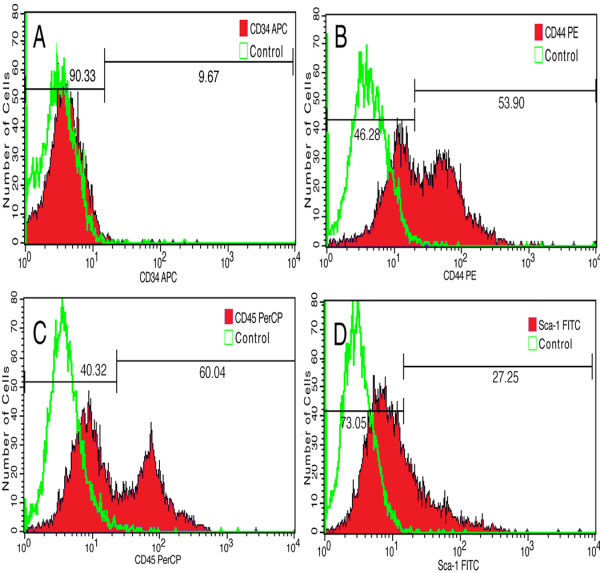
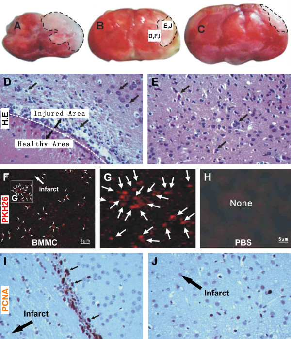
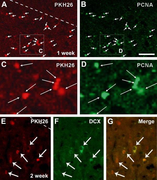
Results: Bone marrow stem cells isolated from donor mice were confirmed by analysis of surface antigen profile, and were pre-labeled with a lipophilic fluorescent dye PKH26, and subsequently transfused into recipient mice with middle cerebral artery coagulation. A large number of PKH26-labeled cells were detected surrounding the infarct site, most of which colocalized with immunolabelings for the proliferating cell nuclear antigen (PCNA) and some also colocalized with the immature neuronal marker doublecortin (DCX) during 1-2 weeks after the bone marrow cells transfusion.
Conclusions: The present study shows that transplanted bone marrow cells largely relocate to the infarct penumbra in ischemic mouse cerebrum. These transplanted bone marrow cells appear to undergo a process of in situ proliferation and develop into putative cortical interneurons during the early phase of experimental vascular injury.
Figures
Similar articles
-
Migration and differentiation of nuclear fluorescence-labeled bone marrow stromal cells after transplantation into cerebral infarct and spinal cord injury in mice.Neuropathology. 2003 Sep;23(3):169-80. doi: 10.1046/j.1440-1789.2003.00496.x. Neuropathology. 2003. PMID: 14570283
-
Human neural progenitor cell engraftment increases neurogenesis and microglial recruitment in the brain of rats with stroke.PLoS One. 2012;7(11):e50444. doi: 10.1371/journal.pone.0050444. Epub 2012 Nov 21. PLoS One. 2012. PMID: 23185625 Free PMC article.
-
In vivo tracking of bone marrow stromal cells transplanted into mice cerebral infarct by fluorescence optical imaging.Brain Res Brain Res Protoc. 2004 Aug;13(3):166-75. doi: 10.1016/j.brainresprot.2004.04.004. Brain Res Brain Res Protoc. 2004. PMID: 15296854
-
[Therapeutic application of cell transplantation and increased neurogenesis in cerebral infarction].Rinsho Shinkeigaku. 2004 Nov;44(11):756-9. Rinsho Shinkeigaku. 2004. PMID: 15651283 Review. Japanese.
-
[Differentiation of adult bone marrow cells into neurons and endothelial cells in rat brain after stroke in the presence of cytokines].Rinsho Shinkeigaku. 2003 Nov;43(11):830-1. Rinsho Shinkeigaku. 2003. PMID: 15152477 Review. Japanese.
Cited by
-
The Use of Stem Cells as a Potential Treatment Method for Selected Neurodegenerative Diseases: Review.Cell Mol Neurobiol. 2023 Aug;43(6):2643-2673. doi: 10.1007/s10571-023-01344-6. Epub 2023 Apr 7. Cell Mol Neurobiol. 2023. PMID: 37027074 Free PMC article. Review.
-
Experimental models of brain ischemia: a review of techniques, magnetic resonance imaging, and investigational cell-based therapies.Front Neurol. 2014 Feb 19;5:19. doi: 10.3389/fneur.2014.00019. eCollection 2014. Front Neurol. 2014. PMID: 24600434 Free PMC article. Review.
-
Granulocyte colony-stimulating factor increases the therapeutic efficacy of bone marrow mononuclear cell transplantation in cerebral ischemia in mice.BMC Neurosci. 2011 Jun 24;12:61. doi: 10.1186/1471-2202-12-61. BMC Neurosci. 2011. PMID: 21699735 Free PMC article.
-
Human Tubal-Derived Mesenchymal Stromal Cells Associated with Low Level Laser Therapy Significantly Reduces Cigarette Smoke-Induced COPD in C57BL/6 mice.PLoS One. 2015 Aug 31;10(8):e0136942. doi: 10.1371/journal.pone.0136942. eCollection 2015. PLoS One. 2015. PMID: 26322981 Free PMC article.
-
Recovery of neurological function of ischemic stroke by application of conditioned medium of bone marrow mesenchymal stem cells derived from normal and cerebral ischemia rats.J Biomed Sci. 2014 Jan 22;21(1):5. doi: 10.1186/1423-0127-21-5. J Biomed Sci. 2014. PMID: 24447306 Free PMC article.
References
MeSH terms
Substances
LinkOut - more resources
Full Text Sources
Medical
Miscellaneous
