. 2010 Oct 25:11:83.
doi: 10.1186/1471-2121-11-83.

The individual-cell-based cryo-chip for the cryopreservation, manipulation and observation of spatially identifiable cells. II: functional activity of cryopreserved cells

Affiliations

The individual-cell-based cryo-chip for the cryopreservation, manipulation and observation of spatially identifiable cells. II: functional activity of cryopreserved cells

Elena Afrimzon et al. BMC Cell Biol. .

Abstract

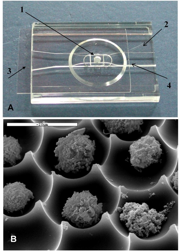
Background: The cryopreservation and thawing processes are known to induce many deleterious effects in cells and might be detrimental to several cell types. There is an inherent variability in cellular responses among cell types and within individual cells of a given population with regard to their ability to endure the freezing and thawing process. The aim of this study was to evaluate the fate of cryopreserved cells within an optical cryo apparatus, the individual-cell-based cryo-chip (i3C), by monitoring several basic cellular functional activities at the resolution of individual cells.

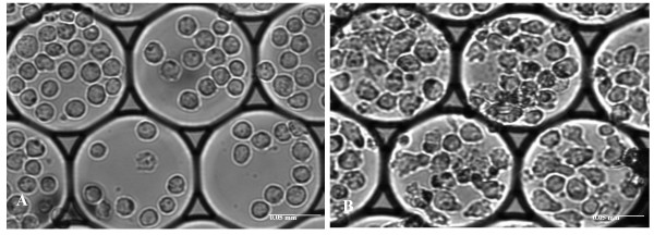
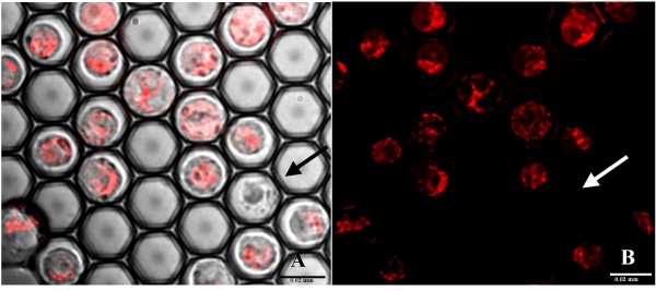
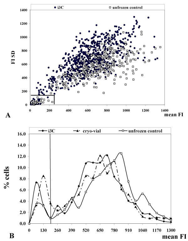
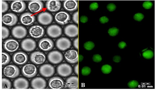
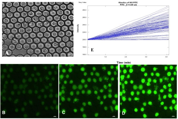
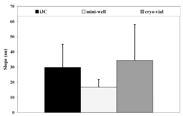
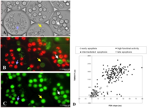
Results: In the present study, U937 cells underwent the freezing and thawing cycle in the i3C device. Then a panel of vital tests was performed, including the number of dead cells (PI staining), apoptotic rate (Annexin V staining), mitochondrial membrane potential (TMRM staining), cytoplasm membrane integrity and intracellular metabolism (FDA staining), as well as post-thawing cell proliferation assays. Cells that underwent the freezing - thawing cycle in i3C devices exhibited the same functional activity as control cells. Moreover, the combination of the multi-parametric analysis at a single cell resolution and the optical and biological features of the device enable an accurate determination of the functional status of individual cells and subsequent retrieval and utilization of the most valuable cells.

Conclusions: The means and methodologies described here enable the freezing and thawing of spatially identifiable cells, as well as the efficient detection of viable, specific, highly biologically active cells for future applications.


Figures

Figure 1
Figure 1
The individual-cell-based cryo-chip (i3C). (A) The 2.5 ml cell chamber (1), whose glass bottom contains the picowells (small opaque region inside the chamber), which are engraved into the i3C plastic body (2). The cover slip (3) is moved left to enable access to the cell chamber opening, through which the cell suspension is loaded. The cover slip is then returned to its position in order to secure the content of the cell chamber when the liquids are poured into the entrance of the open conduit (4), formed by the space between the cover slip and the plastic 'step bridge'. Flow to the left is then established via capillary forces developed between the cover slip, the bridge and the poured liquid. (B) SEM micrograph of U937 cells in their picowells, as they appear at the bottom of the cell chamber.
Figure 2
Figure 2
Cell proliferation after thawing. Bright field images of the cells (objective × 20) in the same individual picowells: A - immediately after the freezing - thawing cycle, B - following 48 hour culturing. Scale bar: 0.05 mm.
Figure 3
Figure 3
Mitochondrial staining of untreated U937 cells by TMRM (200 nM), a potentiometric membrane dye which accumulates in the mitochondria. Images (× 60 magnification): the overlay of transmitted light and fluorescent images (A) and the corresponding fluorescent image (B) are presented. The white arrow indicates a TMRM negative U937 cell, also characterized by its morphological appearance in the transmitted light image. Scale bar: 0.02 mm
Figure 4
Figure 4
Characteristics of the mitochondria in U937 cells after the freezing - thawing cycle (1.5 hours). (A). Scattered diagram of single cells' mean FI (X axis) vs. FI SD (Y axis) for individual cells stained with TMRM in the i3C (1.5 hours after the freezing - thawing cycle) and unfrozen control. Each dot represents an individual cell. (B) Distribution histograms of FI values in individual U937 cells that underwent the freezing - thawing cycle in the i3C, standard cryo-vials and the unfrozen control.
Figure 5
Figure 5
Monitoring U937 cell membrane integrity after the freezing - thawing cycle by FDA staining. The U937 cells were exposed to the freezing - thawing cycle in the i3C. At 3 hours after thawing, FDA solution (1.2 μM) was introduced into the i3C and images were taken 5 minutes after FDA addition. The transmitted image (A) and corresponding fluorescent image (B) are presented. The dead U937 cell unstained by FDA is indicated by the red arrow. Scale bar: 0.01 mm.
Figure 6
Figure 6
Metabolic characteristics of U937 cells that underwent a freezing and thawing cycle in the i3C. At 3 hours after thawing the cells were stained by FDA (1.2 μM) and the kinetics of FI increase were measured. Transmitted light image (A) and the corresponding fluorescence images (B-D) of U937 cells at 1 minute (B), 3 minutes (C) and 5 minutes (D) after FDA addition. Scale bar: 0.01 mm. (E) Intracellular staining rates derived from image analysis of the same U937 cells as in A-D. Each line represents the FI vs. time for an individual cell.
Figure 7
Figure 7
Fluorescein diacetate hydrolysis rates at 19 hours after thawing. For the evaluation of the metabolic recovery after the freezing - thawing cycle, the cells were stained by FDA and the linear slope FI(t) was calculated for each single cell by image analysis. The mean slopes for U937 cell groups that underwent the freezing - thawing cycle in the cryo-devices (at least 4 experiments for each cryo-device) are presented as mean ± SD.
Figure 8
Figure 8
Multi-parametric staining of cryopreserved U937 cells. U937 cells were loaded into 100 μm i3C and underwent a freezing - thawing cycle. After 24 hours of post-thawing culture, the U937 cells were stained sequentially first by TMRM and Annexin V and next by FDA and PI. Transmitted light (A) and fluorescent images of U937 cells stained with TMRM and Annexin V (B), and FDA (C) are presented. Arrows indicate different cell statuses: Blue - high functional activity - morphologically intact, highly TMRM and FDA positive, Annexin V negative; Yellow - early apoptosis - a morphologically changed cell with a slightly condensed nucleus, exhibiting low TMRM staining, FDA positive staining and strong Annexin V staining; White - late apoptosis - a morphologically changed cell with nucleus defragmentation and pronounced Annexin V positive staining, TMRM and FDA negative staining. Scale bar: 0.05 mm. (D) Scatter diagram of individual TMRM (FI) values vs. esterase enzymatic activity values (slopes), measured in post-thawed U937 cells 24 hours after cell thawing. Note the detection of 4 cell subgroups based on their functional activity parameters.

References

    1. Lermen D, Blomeke B, Rowne R, Clarke A, Dyce PW, Fixemer T, Fuhr G, Holt WV, Ewgenow KJ, Lloyd RE, Lotters S, Paulus M, Reid GMc, Rapoport DH, Rawson D, Ringleb J, Ryder OA, Sporl G, Schmitt T, Veith M, Muller P. Cryobanking of viable biomaterials: implementation of new strategies for conservation purposes. Mol Ecol. 2009;18:1030–1033. doi: 10.1111/j.1365-294X.2008.04062.x. - DOI - PubMed
    1. Rott NN. The creation of genetic cryobanks and the use of the methods of developmental biology as a means for preserving rare animal species. II. The obtaining and cryopreservation of the embryos of wild mammals. Ontogenez. 1996;27:245–255. - PubMed
    1. Grout BW. Cryopreservation of plant cell suspensions. Methods Mol Biol. 2007;368:153–161. full_text. - PubMed
    1. Van Hoof D, D'Amour KA, German MS. Derivation of insulin-producing cells from human embryonic stem cells. Stem Cell Res. 2009;3:73–87. doi: 10.1016/j.scr.2009.08.003. - DOI - PubMed
    1. McCall MD, Toso C, Baetge EE, Shapiro AM. Are stem cells a cure for diabetes? Clin Sci. 2009;118:87–97. doi: 10.1042/CS20090072. - DOI - PubMed

Publication types