. 2010 Oct 25:7:51.
doi: 10.1186/1476-9255-7-51.

Defining human mesenchymal stem cell efficacy in vivo

Affiliations

Defining human mesenchymal stem cell efficacy in vivo

Tracey L Bonfield et al. J Inflamm (Lond). .

Abstract

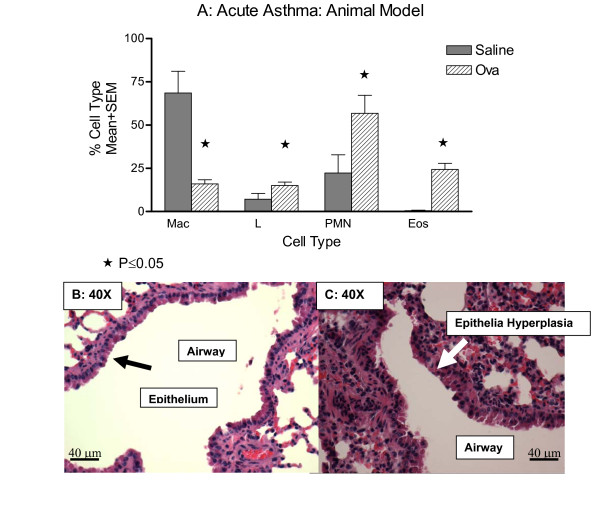
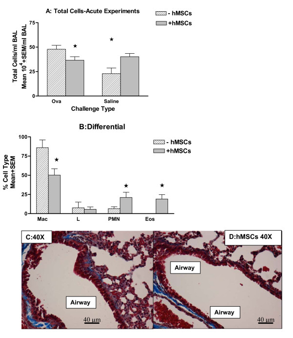
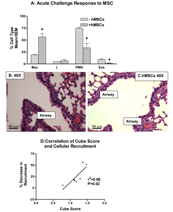
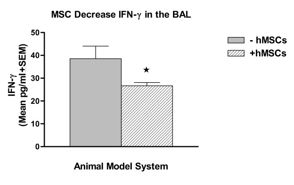
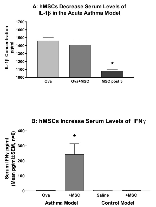
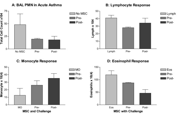
Allogeneic human mesenchymal stem cells (hMSCs) can suppress graft versus host disease (GvHD) and have profound anti-inflammatory and regenerative capacity in stroke, infarct, spinal cord injury, meniscus regeneration, tendinitis, acute renal failure, and heart disease in human and animal models of disease. There is significant clinical hMSC variability in efficacy and the ultimate response in vivo. The challenge in hMSC based therapy is defining the efficacy of hMSC in vivo. Models which may provide insight into hMSC bioactivity in vivo would provide a means to distinguish hMSCs for clinical utility. hMSC function has been described as both regenerative and trophic through the production of bioactive factors. The regenerative component involves the multi-potentiality of hMSC progenitor differentiation. The secreted factors generated by the hMSCs are milieu and injury specific providing unique niches for responses in vivo. These bioactive factors are anti-scarring, angiogenic, anti-apoptotic as well as regenerative. Further, from an immunological standpoint, hMSC's can avoid host immune response, providing xenographic applications. To study the in vivo immuno-regulatory effectiveness of hMSCs, we used the ovalbumin challenge model of acute asthma. This is a quick 3 week in vivo pulmonary inflammation model with readily accessible ways of measuring effectiveness of hMSCs. Our data show that there is a direct correlation between the traditional ceramic cube score to hMSCs attenuation of cellular recruitment due to ovalbumin challenge. The results from these studies verify the in vivo immuno-modulator effectiveness of hMSCs and support the potential use of the ovalbumin model as an in vivo model of hMSC potency and efficacy. Our data also support future directions toward exploring hMSCs as an alternative therapeutic for the treatment of airway inflammation associated with asthma.


Figures

Figure 1
Figure 1
Establishing the Acute Model of Murine Asthma. Mice were sensitized with ovalbumin, rested for 14 days and then challenged daily with ovalbumin or sham PBS for 5 days. Mice were sacrificed and the lungs were processed for inflammation with BAL (1A) or histology without BAL (1B saline-challenged (40×) versus 1C ovalbumin-challenged (40×)). Histology is representative of 5 different experiments with 6-8 mice in each group. Inflammation is representative of 5 different experiments with n = 4-6 for each group. Ovalbumin challenged mice had a significant increase in inflammatory cells (n = 4, p = 0.04).
Figure 2
Figure 2
Dil labeled hMSCs can be Localized in the Murine Lung. Sensitized and challenged mice from the acute asthma group were given hMSCs 5 days prior to euthanisia. The hMSCs were labeled with the vital dye, Dil. The animals were assessed for the presence of Dil fluorescent hMSCs. Highly positive hMSCs (white arrows) were observed in the lung tissue of mice #11 and #26,.
Figure 3
Figure 3
Xenographic Effect of hMSCs on Total Cell Recruitment. Animals were sensitized with ovalbumin and challenged with saline followed by BAL. The total differential cell count between saline-challenged mice and naive controls was negligible (data not shown). hMSC exposure decreased the total cell count in the BAL of the ovalbumin-challenged mice (Figure 3A, p = 0.03, n = 5), but increased the total cell count in the saline-challenged mice (Figure 3A, p < 0.05 for the saline control versus saline control with husks). Further, the hMSCs altered the inflammatory phenotype of the saline control animal (3B, n = 4, p < 0.05) suggesting that the hMSCs can induced a cellular response in the absence of ovalbumin-induced inflammation in the context of an acute response. The change in the inflammatory status did not impact the immediate histology of the acute treated model (3C:40× and 3D:hMSC, 40×). Histology is representative of 5 different experiments with n = 6-8 in each group.
Figure 4
Figure 4
hMSCs Decrease Acute Inflammation in the Acute Asthma Lung Model. hMSCs (106/100 ul injected) were given by tail vein injection at day 14 post-sensitization. Mice were evaluated after 5 days of challenge for inflammation. Concurrent mice were evaluated specifically for histology (Figure 4B and 4C). Treatment of the acute asthma mice with MSCs resulted in increased production of macrophages and decreased production of neutrophils and eosinophils. Histologically, the epithelial lining of the bronchiolar airways appears to have less thickening and less surrounding mucus (4C: 40×) when compared with animals not treated with hMSCs (4B: 40×). Histology is representative of 5 different experiments with 6-8 mice in each group. Inflammation is representative of 5 different experiments with n = 4-6 for each group. To determine the relationship between response to hMSCs and the differentiation of the hMSCs, the percent change in total cellular recruitment post-hMSC treatment was plotted against the cube score values in Table I (4D). Cube score values statistically correlated to percent decrease in cellular recruitment (decrease in inflammation) with r2 = 0.68, p = 0.02, n = 7 different cube scores on n = 7 different hMSCs in 7 different ovalbumin challenged asthma experiments.
Figure 5
Figure 5
hMSCs Decrease IFNγ in Acute Asthma. BAL fluid obtained from mice modeled for acute asthma had significantly less IFNγ when treated with hMSCs in vivo (n = 4, p < 0.05).
Figure 6
Figure 6
Serum IL-1β Decreases with hMSC Treatment. Cardiac puncture was done to obtain adequate serum samples from mice with and without treatment with hMSCs. The acute asthma mice had significantly elevated levels of systemic IL-1β (6A, n = 8, p < 0.001). Systemic IL-1β concentrations were altered by hMSCs in the acute asthma model when hMSCs were given three days after challenge (peak inflammation). hMSCs significantly decreased the level of systemic IL-1β (n = 2, p = 0.05). Systemic IFNγ concentrations were increased in the acute asthma mice after hMSC treatment (6B, n = 4, p = 0.043). This was dependent on the presence of in vivo inflammation, since saline animals given hMSCs had no detectable serum IFNγ.
Figure 7
Figure 7
Lung Inflammation Determines Response to hMSCs. BAL fluid was obtained from mice treated with hMSCs just prior to challenge and initiation of inflammation (pre-) or 3 days after initiation of inflammation (post-). BAL differentials were obtained from each animal using cytospins and Wright-Giemsa stain. Neutrophil counts are in panel A, lymphocytes in panel B, monocytes in panel C and eosinophils in panel D. The first bar represents the differential in the model without hMSCs. No difference was observed in levels of neutrophils or lymphocytes after hMSC treatment. Pre-challenged mice had a decrease in eosinophils (panel D, n = 4, p = 0.05) compared to control mice. This decrease in eosinophils was amplified at post-treatment with hMSCs. Additionally, post-treatment mice had a significant increase in localized levels of monocyte/macrophages (panel C, n = 2, p = 0.05, 4 animals used in each group for each experiment).

References

    1. Caplan AI. All MSCs are pericytes? Cell Stem Cell. 2008;3:229–230. doi: 10.1016/j.stem.2008.08.008. - DOI - PubMed
    1. da Silva ML, Caplan AI, Nardi NB. In search of the in vivo identity of mesenchymal stem cells. Stem Cells. 2008;26:2287–2299. doi: 10.1634/stemcells.2007-1122. - DOI - PubMed
    1. Caplan AI, Bruder SP. Mesenchymal stem cells: building blocks for molecular medicine in the 21st century. Trends Mol Med. 2001;7:259–264. doi: 10.1016/S1471-4914(01)02016-0. - DOI - PubMed
    1. Dominici M, Le Blanc K, Mueller I, Slaper-Cortenbach I, Marini F, Krause D, Deans R, Keating A, Prockop D, Horwitz E. Minimal criteria for defining multipotent mesenchymal stromal cells. The International Society for Cellular Therapy position statement. Cytotherapy. 2006;8:315–317. doi: 10.1080/14653240600855905. - DOI - PubMed
    1. Chamberlain G, Fox J, Ashton B, Middleton J. Concise review: mesenchymal stem cells: their phenotype, differentiation capacity, immunological features, and potential for homing. Stem Cells. 2007;25:2739–2749. doi: 10.1634/stemcells.2007-0197. - DOI - PubMed

LinkOut - more resources