. 2011 Jan 1;50(1):27-36.
doi: 10.1016/j.freeradbiomed.2010.10.702. Epub 2010 Oct 23.

Selective macrophage ascorbate deficiency suppresses early atherosclerosis

Affiliations

Selective macrophage ascorbate deficiency suppresses early atherosclerosis

Vladimir R Babaev et al. Free Radic Biol Med. .

Abstract

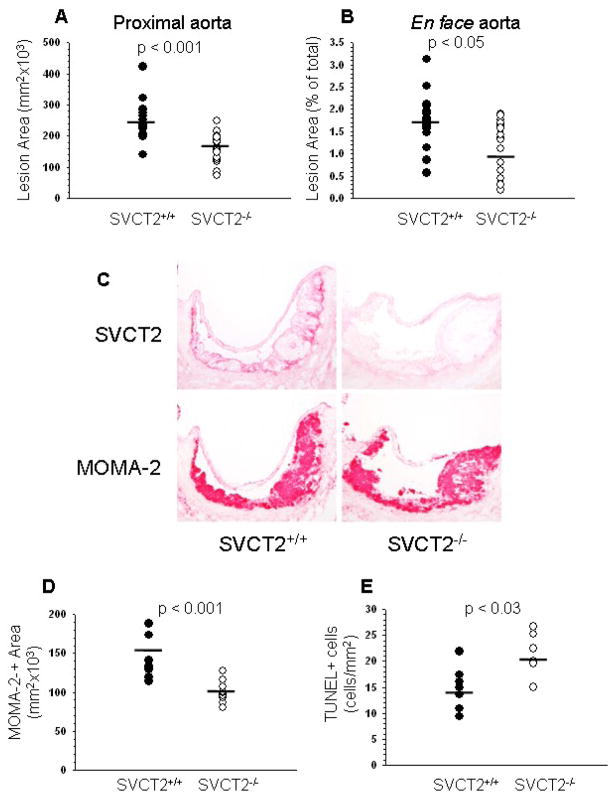
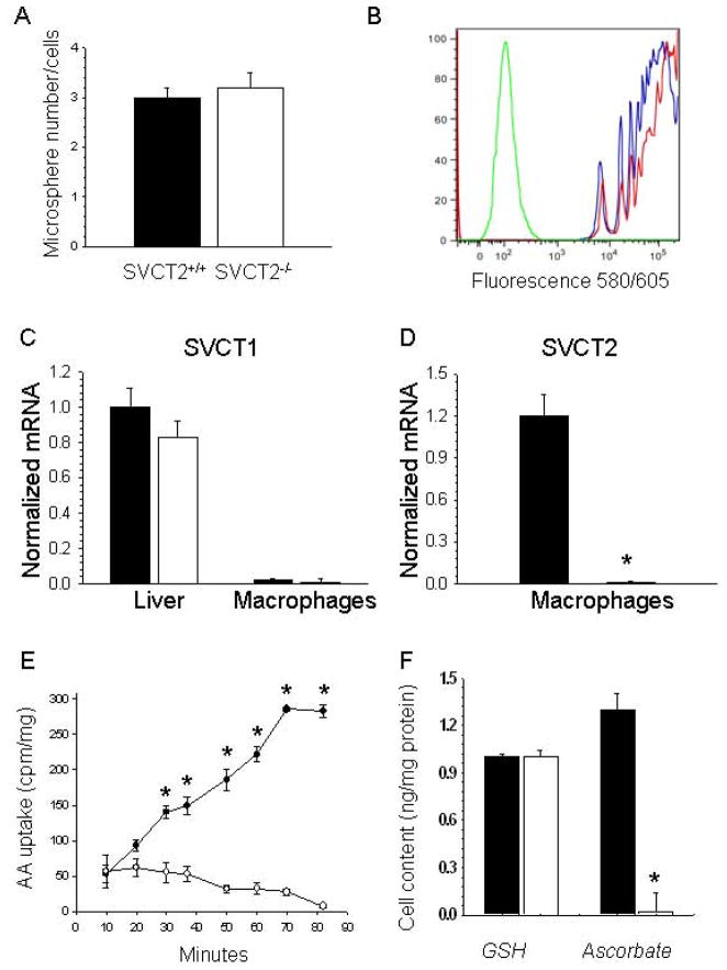
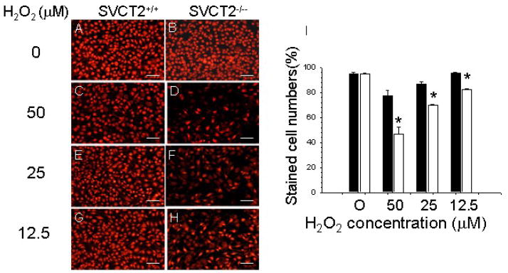
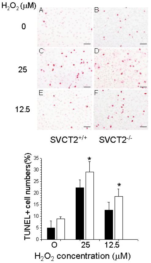
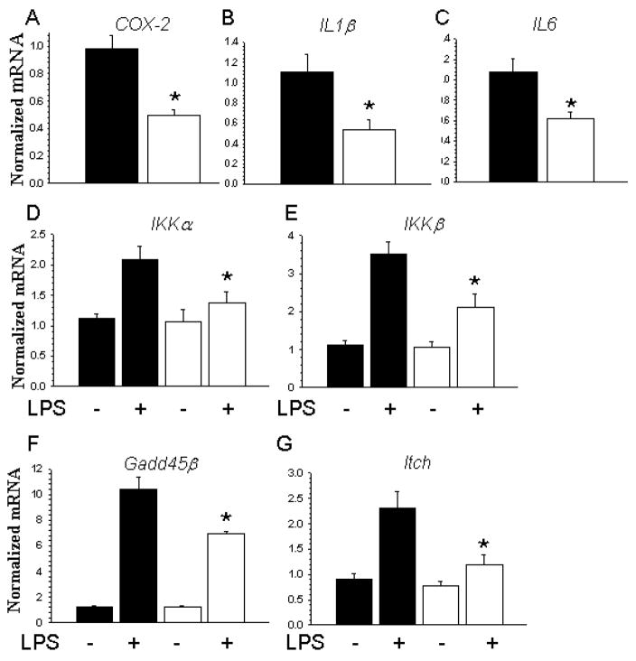
To test whether severe ascorbic acid deficiency in macrophages affects progression of early atherosclerosis, we used fetal liver cell transplantation to generate atherosclerosis-prone apolipoprotein E-deficient (apoE(-/-)) mice that selectively lacked the ascorbate transporter (SVCT2) in hematopoietic cells, including macrophages. After 13 weeks of chow diet, apoE(-/-) mice lacking the SVCT2 in macrophages had surprisingly less aortic atherosclerosis, decreased lesion macrophage numbers, and increased macrophage apoptosis compared to control-transplanted mice. Serum lipid levels were similar in both groups. Peritoneal macrophages lacking the SVCT2 had undetectable ascorbate; increased susceptibility to H(2)O(2)-induced mitochondrial dysfunction and apoptosis; decreased expression of genes for COX-2, IL1β, and IL6; and decreased lipopolysaccharide-stimulated NF-κB and antiapoptotic gene expression. These changes were associated with decreased expression of both the receptor for advanced glycation end products and HIF-1α, either or both of which could have been the proximal cause of decreased macrophage activation and apoptosis in ascorbate-deficient macrophages.


Figures

Figure 1
Figure 1
Atherosclerosis, SVCT2 expression and apoptosis in mouse atherosclerotic lesions. (A, B) Image analysis data obtained from serial sections of aortic sinus (A) and en face aorta (B) of mice reconstituted with apoE−/− FLCs either expressing the SVCT2 (SVCT2+/+, n = 18) or lacking it (SVCT2−/−, n = 19). (C) Immunostaining of sequential random aortic sinus sections for the SVCT2 (top panels) and macrophages (MOMA-2, bottom panels) in apoE−/− mice transplanted with SVCT2+/+ or SVCT2−/− FLCs. (D) Image analysis of MOMA-2 immunostaining from mice transplanted with SVCT2+/+ FLCs or mice transplanted with SVCT2−/− FLCs. (E) Image analysis of TUNEL-positive cells in proximal aortic lesions from mice transplanted with SVCT2+/+ FLCs or from mice transplanted with SVCT2−/− FLCs. In A, B, D and E, bars indicate mean values.
Figure 2
Figure 2
Macrophage phagocytosis and ascorbate transporter expression and function. Liver tissue and peritoneal macrophages were isolated from apoE−/− mice reconstituted either with SVCT2+/+ (■) or SVCT2−/− (□) FLCs. Phagocytosis of fluorescent microspheres was allowed to continue for 1 hour at 37°C, followed either by counting of macrophage bead content (A) or by flow cytometry (B). For (B), the green line represents control non-phagocytosing macrophages, the red line macrophages from mice transplanted with SVCT2−/− FLCs, and the blue line macrophages from mice that received SVCT2+/+ FLCs. Gene expression levels of SVCT1 (C) and SVCT2 (D) were measured in liver and macrophages by real-time PCR. Graphs show results from 3 mice per group, with an asterisk indicating p < 0.05 compared to the control group. Radiolabeled ascorbate uptake over 90 min was measured in peritoneal macrophages in 4 experiments, with an asterisk indicating p < 0.05 compared to time zero (E). Macrophage ascorbate and GSH were measured in macrophages from each group that were cultured for 2 days after harvesting (F). Results are shown from 4 experiments, with an asterisk indicating p < 0.05 compared to control ascorbate.
Figure 3
Figure 3
SVCT2 deficiency impairs mitochondrial function in response to H2O2. (A–H) Staining by MitoTracker red of mitochondria in SVCT2+/+ and SVCT2−/− macrophages treated with the indicated amounts of H2O2. The scale bars represent 50 μm. (I) Mitotracker red staining of SVCT2+/+ (■) and SVCT2−/− (□) macrophages from 3 mice in each group, with an asterisk indicating p < 0.05 compared to SVCT2+/+ macrophages at each H2O2 concentration.
Figure 4
Figure 4
SVCT2 deficiency increases TUNEL+ cell numbers in response to H2O2. Top panel: TUNEL staining of SVCT2+/+ and SVCT2−/− macrophages and treated with increasing concentrations of H2O2 (A–F). The scale bars represent 50 μm. Bottom Panel: Percent of TUNEL+ macrophages from 3 mice for SVCT2+/+ (■) and SVCT2−/− (□) macrophages. An asterisk indicates p < 0.05 compared to SVCT2+/+ macrophages.
Figure 5
Figure 5
Lack of SVCT2 suppresses RAGE gene and protein expression in peritoneal macrophages and aortic sinus lesions. RAGE mRNA (A) and protein (B) levels were measured in untreated SVCT2+/+ (■) and SVCT2−/− (□) macrophages from 3 mice in each group. An asterisk indicates p < 0.05 compared to control macrophages. (C) Representative immunostaining for RAGE (top panels) and macrophages (bottom panels) of cross sections of aortic sinus of mice reconstituted with SVCT2+/+/apoE−/− (left-hand panels) and SVCT2−/−/apoE−/− (right-hand panels) FLCs.
Figure 6
Figure 6
SVCT2 deficiency in macrophages suppresses NF-κB-related and anti-apoptotic genes. (A–C) Basal expression of mRNA for inflammation-related genes (COX-2, IL1β and IL6) in untreated SVCT2+/+ (■) and SVCT2−/− (□) macrophages from 3 mice in each group. Data are shown as mean ± SEM, with an asterisk indicating p < 0.05 compared to the control group. (D–G) SVCT2+/+ (■) and SVCT2−/− (□) macrophages from 3 mice in each group were treated with 50 nM LPS for 5 hours, followed by measurement of mRNA for NF-κB-related (IKKα, IKKβ) and anti-apoptotic (Gadd45β and Itch) genes. An asterisk indicates p < 0.05 compared to control LPS treatment for the respective group.
Figure 7
Figure 7
Macrophage SVCT2 deficiency inhibits expression of HIF-1α. HIF-1α protein expression is shown for macrophages either lacking or expressing the SVCT2 from 5 mice (A). In (B) the results are shown relative to actin expression, with an asterisk indicating p < 0.05 compared to control macrophages.

References

    1. Ross R. Atherosclerosis--an inflammatory disease. N Engl J Med. 1999;340:115–26. - PubMed
    1. Linton MF, Fazio S. Macrophages, inflammation, and atherosclerosis. Int J Obes Relat Metab Disord. 2003;27(Suppl 3):S35–S40. - PubMed
    1. Cook NR, Albert CM, Gaziano JM, Zaharris E, MacFadyen J, Danielson E, et al. A randomized factorial trial of vitamins C and E and beta carotene in the secondary prevention of cardiovascular events in women: results from the Women’s Antioxidant Cardiovascular Study. Arch Intern Med. 2007;167:1610–8. - PMC - PubMed
    1. Heart Protection Study Collaborative Group. MRC/BHF Heart Protection Study of antioxidant vitamin supplementation in 20,536 high-risk individuals: a randomised placebo-controlled trial. Lancet. 2002;360:23–33. - PubMed
    1. Sesso HD, Buring JE, Christen WG, Kurth T, Belanger C, MacFadyen J, et al. Vitamins E and C in the prevention of cardiovascular disease in men: the Physicians’ Health Study II randomized controlled trial. JAMA. 2008;300:2123–33. - PMC - PubMed

Publication types

MeSH terms