

Increased interstitial white matter neuron density in the dorsolateral prefrontal cortex of people with schizophrenia
- PMID: 20974464
- PMCID: PMC3005941
- DOI: 10.1016/j.biopsych.2010.08.020
Increased interstitial white matter neuron density in the dorsolateral prefrontal cortex of people with schizophrenia
Abstract
Background: Interstitial white matter neurons (IWMNs) may reflect immature neurons that migrate tangentially to the neocortex from the ganglionic eminence to form cortical interneurons. Alterations of interneuron markers have been detected in gray matter of dorsolateral prefrontal cortex in schizophrenia, and IWMNs are also reported to be altered in schizophrenia. In this study, we considered whether a potential link exists between these two pathological findings.
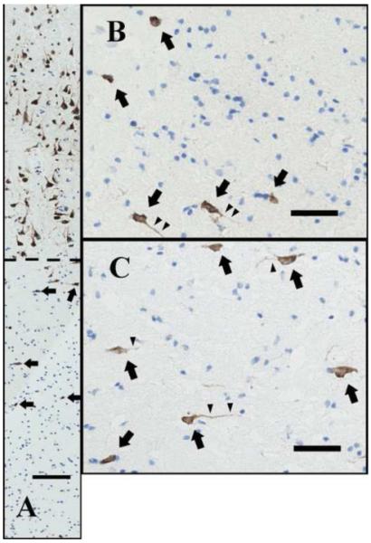
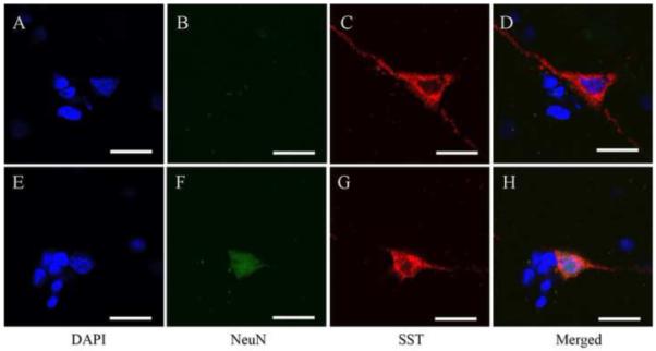
Methods: From a cohort of 29 schizophrenia subjects and 37 control subjects, IWMN densities were determined in the dorsolateral prefrontal cortex by counting neuronal nuclear antigen (NeuN) and somatostatin (SST)-positive cells. Double-label immunofluorescence was carried out to determine the overlap between SST+/NeuN+ and SST+/neuropeptide Y + neurons.
Results: We found that density of NeuN + IWMNs in superficial white matter is significantly increased in schizophrenia subjects compared with control subjects. There was a significant negative correlation between SST mRNA expression in gray matter and NeuN + IWMN density. In schizophrenic patients with increased NeuN IWMN density, the density of SST-expressing neurons in white matter was also higher compared with control subjects. A subpopulation of SST immunopositive cells also show coexpression of neuropeptide Y.
Conclusions: Our study confirmed previous results indicating that the density of NeuN + IWMNs is increased in superficial white matter in schizophrenia. We provide the first evidence that increased IWMN density correlates with a gray matter interneuron deficit, suggesting that migration of interneurons from white matter to the cortex may be deficient in some patients with schizophrenia, consistent with an interneuron deficit in schizophrenia.
Copyright © 2011 Society of Biological Psychiatry. Published by Elsevier Inc. All rights reserved.
Figures


Similar articles
-
Higher gamma-aminobutyric acid neuron density in the white matter of orbital frontal cortex in schizophrenia.Biol Psychiatry. 2012 Nov 1;72(9):725-33. doi: 10.1016/j.biopsych.2012.06.021. Epub 2012 Jul 25. Biol Psychiatry. 2012. PMID: 22841514
-
Increased white matter neuron density in a rat model of maternal immune activation - Implications for schizophrenia.Prog Neuropsychopharmacol Biol Psychiatry. 2016 Feb 4;65:118-26. doi: 10.1016/j.pnpbp.2015.09.006. Epub 2015 Sep 15. Prog Neuropsychopharmacol Biol Psychiatry. 2016. PMID: 26385575
-
Cingulate white matter neurons in schizophrenia and bipolar disorder.Biol Psychiatry. 2009 Sep 1;66(5):486-93. doi: 10.1016/j.biopsych.2009.04.032. Epub 2009 Jun 25. Biol Psychiatry. 2009. PMID: 19559403 Free PMC article.
-
White matter neuron biology and neuropathology in schizophrenia.NPJ Schizophr. 2019 Jul 8;5(1):10. doi: 10.1038/s41537-019-0078-8. NPJ Schizophr. 2019. PMID: 31285426 Free PMC article. Review.
-
White matter neuron alterations in schizophrenia and related disorders.Int J Dev Neurosci. 2011 May;29(3):325-34. doi: 10.1016/j.ijdevneu.2010.07.236. Epub 2010 Aug 4. Int J Dev Neurosci. 2011. PMID: 20691252 Free PMC article. Review.
Cited by
-
Increased density of prohibitin-immunoreactive oligodendrocytes in the dorsolateral prefrontal white matter of subjects with schizophrenia suggests extraneuronal roles for the protein in the disease.Neuromolecular Med. 2012 Dec;14(4):270-80. doi: 10.1007/s12017-012-8185-y. Epub 2012 Jun 19. Neuromolecular Med. 2012. PMID: 22711522
-
Nitric oxide synthase 1 adaptor protein, a protein implicated in schizophrenia, controls radial migration of cortical neurons.Biol Psychiatry. 2015 Jun 1;77(11):969-78. doi: 10.1016/j.biopsych.2014.10.016. Epub 2014 Oct 30. Biol Psychiatry. 2015. PMID: 25542305 Free PMC article.
-
Transcriptional co-regulation of neuronal migration and laminar identity in the neocortex.Development. 2012 May;139(9):1535-46. doi: 10.1242/dev.069963. Development. 2012. PMID: 22492350 Free PMC article. Review.
-
Aggrecan and chondroitin-6-sulfate abnormalities in schizophrenia and bipolar disorder: a postmortem study on the amygdala.Transl Psychiatry. 2015 Jan 20;5(1):e496. doi: 10.1038/tp.2014.128. Transl Psychiatry. 2015. PMID: 25603412 Free PMC article.
-
Association of serum VEGF levels with prefrontal cortex volume in schizophrenia.Mol Psychiatry. 2016 May;21(5):686-92. doi: 10.1038/mp.2015.96. Epub 2015 Jul 14. Mol Psychiatry. 2016. PMID: 26169975
References
-
- Harrison PJ. Schizophrenia: a disorder of neurodevelopment? Curr Opin Neurobiol. 1997;7:285–289. - PubMed
-
- Lewis DA, Levitt P. Schizophrenia as a disorder of neurodevelopment. Annu Rev Neurosci. 2002;25:409–432. - PubMed
-
- Weickert CS, Weinberger DR. A candidate molecule approach to defining developmental pathology in schizophrenia. Schizophr Bull. 1998;24:303–316. - PubMed
-
- Weickert CS, Kleinman JE. The neuroanatomy and neurochemistry of schizophrenia. Psychiatr Clin North Am. 1998;21:57–75. - PubMed
-
- Lewis DA, Pierri JN, Volk DW, Melchitzky DS, Woo TU. Altered GABA neurotransmission and prefrontal cortical dysfunction in schizophrenia. Biol Psychiatry. 1999;46:616–626. - PubMed
Publication types
MeSH terms
Substances
Grants and funding
LinkOut - more resources
Full Text Sources
Medical
