. 2011 Mar;39(4):1310-25.
doi: 10.1093/nar/gkq933. Epub 2010 Oct 25.

The transcriptional co-activator LEDGF/p75 displays a dynamic scan-and-lock mechanism for chromatin tethering

Affiliations

The transcriptional co-activator LEDGF/p75 displays a dynamic scan-and-lock mechanism for chromatin tethering

Jelle Hendrix et al. Nucleic Acids Res. 2011 Mar.

Abstract

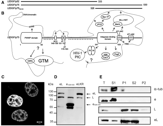
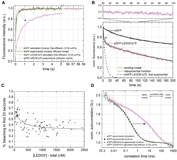
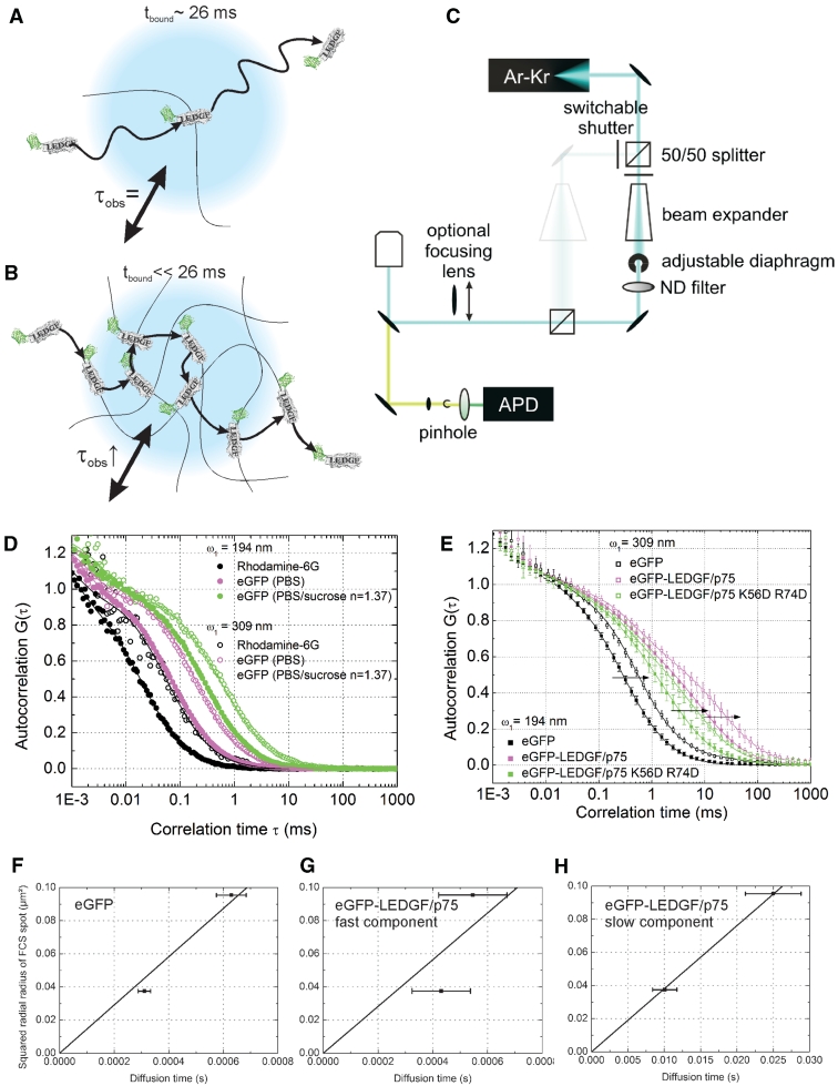
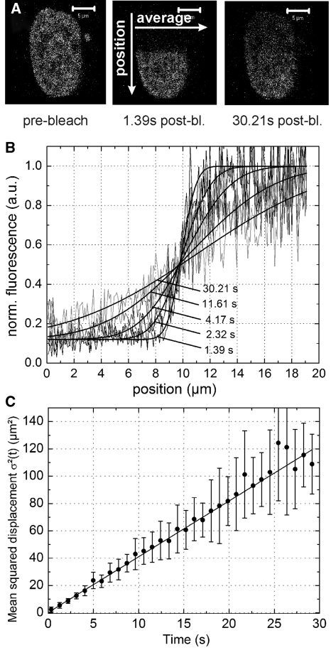
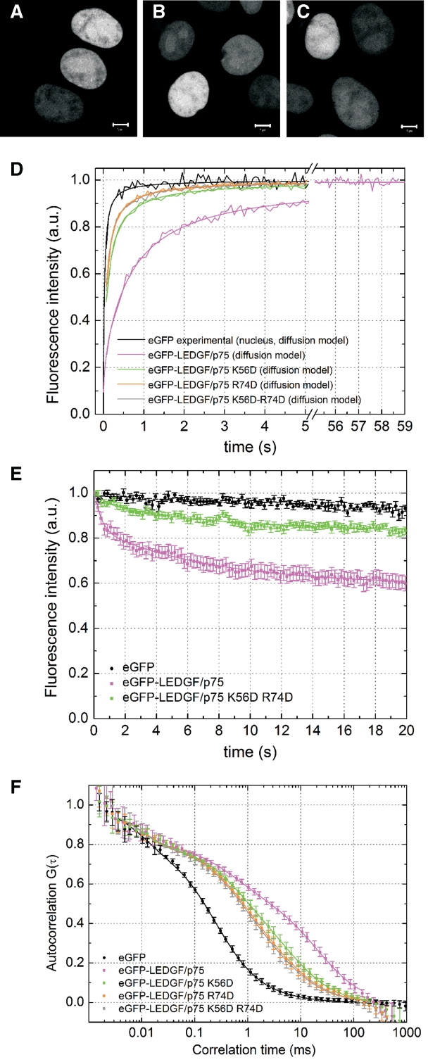
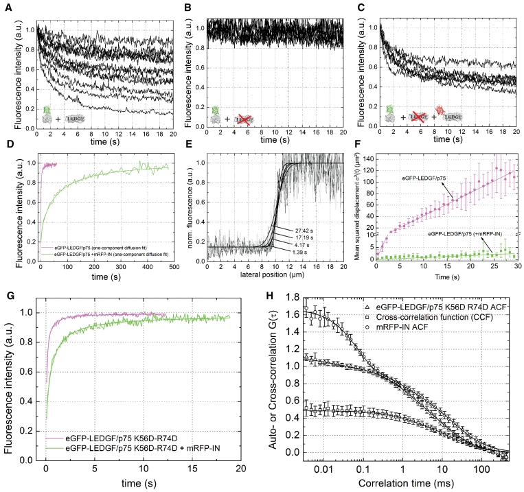
Nearly all cellular and disease related functions of the transcriptional co-activator lens epithelium-derived growth factor (LEDGF/p75) involve tethering of interaction partners to chromatin via its conserved integrase binding domain (IBD), but little is known about the mechanism of in vivo chromatin binding and tethering. In this work we studied LEDGF/p75 in real-time in living HeLa cells combining different quantitative fluorescence techniques: spot fluorescence recovery after photobleaching (sFRAP) and half-nucleus fluorescence recovery after photobleaching (hnFRAP), continuous photobleaching, fluorescence correlation spectroscopy (FCS) and an improved FCS method to study diffusion dependence of chromatin binding, tunable focus FCS. LEDGF/p75 moves about in nuclei of living cells in a chromatin hopping/scanning mode typical for transcription factors. The PWWP domain of LEDGF/p75 is necessary, but not sufficient for in vivo chromatin binding. After interaction with HIV-1 integrase via its IBD, a general protein-protein interaction motif, kinetics of LEDGF/p75 shift to 75-fold larger affinity for chromatin. The PWWP is crucial for locking the complex on chromatin. We propose a scan-and-lock model for LEDGF/p75, unifying paradoxical notions of transcriptional co-activation and lentiviral integration targeting.


Figures

Figure 1.
Figure 1.
The primary structure and interactome of LEDGF/p75. (A) Schematic representation of LEDGF/p75, its alternative splice variant LEDGF/p52 (13) and a deletion mutant, LEDGF/p75326–530 (61). (B) Primary structure and interactome of LEDGF/p75. Important domains of LEDGF/p75, interacting proteins or DNA-sequences are indicated. NLS, nuclear localization signal; AT, AT-hook domains; HTH, predicted Helix-Turn-Helix motifs; GSRs, gene specific regulators; GTM, general transcription machinery; STRE, stress-related regulatory element; HIV-1 PIC, pre-integration complex; IN, HIV-1 integrase; Plusses, positively charged regions in the p52 part of LEDGF/p75. (C) Confocal fluorescence image of HeLa cells expressing eGFP-LEDGF/p75. Scale bar = 5 µm. (D) Western blot with an anti-LEDGF/p75 antibody of HeLa cells transiently expressing eGFP-fusions. eL, eGFP-LEDGF/p75; e326-530, eGFP-LEDGF/p75326-530; eLKR, K56D-R74D; L, endogenous LEDGF/p75. (E) Cellular fractionation assay of HeLa cells transiently expressing eGFP-fusions. Western blot of different fractions is shown using antibodies to indicated proteins. T, total cell lysate; S1, Triton-soluble cellular fraction; P1, Triton-insoluble cellular fraction; S2, DNase/(NH4)2SO4-soluble cellular fraction; P2, DNase/(NH4)2SO4-insoluble cellular fraction.
Figure 2.
Figure 2.
Dynamic subpopulations of LEDGF/p75 revealed by sFRAP, CP and FCS. (A) sFRAP experiment of eGFP and eGFP-LEDGF/p75 in living HeLa cells. The diffusion coefficient we calculated for eGFP after fitting with a pure diffusion model was only 10.28 µm2/s (Table 1). For eGFP the observed recovery was lower than expected (solid orange curve) because of unavoidable recovery-while-photobleaching, which occurred because the diffusion time of eGFP (tdiff = 97 ms) was too small with respect to the acquisitioning time of the microscope (65 ms per iteration on our setup) (40). For eGFP-LEDGF/p75, the arrow indicates the decrease in mobility of eGFP-LEDGF/p75 due to chromatin interactions. DeGFP = 33 µm2/s is the D we measured with FCS, DeGFP-LEDGF/p75 = 22 µm2/s is calculated from DeGFP with Equation 8. See also Supplementary Figure S1. (B) CP experiment of eGFP and eGFP-LEDGF/p75. Upper panel is a 80–100-s zoom. (C) Concentration dependence of the amount of photobleaching of eGFP-LEDGF/p75 during the first 20 s. Solid line = fit with Equation 6. (D) FCS experiment of eGFP and eGFP-LEDGF/p75. Solid lines = two-component normal diffusion model. See also Supplementary Figure S2. Error bars = SD.
Figure 3.
Figure 3.
TFFCS shows fast chromatin binding of LEDGF/p75. (A) The observed slow diffusion time (τobs = 25.9 ms, D = 0.5 µm2/s) is the binding time of LEDGF/p75 with chromatin, tbound = 1/koff. In this case τobs does not scale with the laser focus diameter, indicated in blue. (B) LEDGF/p75 continuously associates with and dissociates from the chromatin while diffusing through the measurement spot. The τobs scales with the laser focus diameter. (C) Schematic representation of the home built TFFCS set-up. The beam expander and diaphragm are used to vary the diameter of the collimated laser beam. ND, neutral density attenuation filter; APD, avalanche photodiode. The diameter of the diaphragm is controlled by measuring the power of the excitation light passing through it. Higher power through the diaphragm means a broader beam will enter the objective back aperture. The objective focuses a broader beam in a smaller focal point, since the beam diameter determines the effective numerical aperture of the objective (NA = n × sinα with n the refractive index and α the angular aperture). For further details, see the ‘Materials and Methods’ section. (D) In vitro TFFCS measurements of rhodamine 6G in water (black), eGFP in PBS (magenta) and PBS/sucrose with a viscosity and refractive index similar to that of the intracellular environment (n = 1.37) (green). ω1: radial radius of the excitation spot. (E) Intracellular TFFCS measurements of eGFP (black), eGFP-LEDGF/p75 (magenta) and eGFP-LEDGF/p75 K56D-R74D (green). Error bars = SD. (FH) Plot of the diffusion time from the experimental ACF versus the squared radial radius of the confocal excitation spot for (F) eGFP, (G) the fast and (H) the slow component of eGFP-LEDGF/p75.
Figure 4.
Figure 4.
LEDGF/p75 shows normal diffusion dependent dynamics. (A) Illustration of half-nucleus FRAP. Half of the nucleus is photobleached and recovery perpendicular to the bleach border is monitored versus time. (B) Nuclear intensity profile versus time along the arrow designated ‘position’ in (A) Solid lines = Equation 10. (C) Mean squared displacement – time plot. Error bar = SD. Solid line = Equation 11.
Figure 5.
Figure 5.
The PWWP domain of LEDGF/p75 contributes to high affinity chromatin binding and is crucial for chromatin tethering of HIV-1 integrase. (A–C) Confocal fluorescence images of HeLa cells expressing (A) eGFP-LEDGF/p75 K56D, (B) R74D and (C) K56D-R74D. (D) sFRAP experiment. (E) CP experiment of eGFP-LEDGF/p75 K56D-R74D. The CP curves of eGFP and eGFP-LEDGF/p75 are shown as a reference. (F) FCS experiment of eGFP-LEDGF/p75 mutants. The ACFs of eGFP and eGFP-LEDGF/p75 are shown as a reference. Error bars = SD.
Figure 6.
Figure 6.
LEDGF/p75-PWWP locks IN-LEDGF/p75 on chromatin. (A–C) CP experiment of eGFP-IN in (A) wild-type HeLa cells, (B) HeLa-p75KD cells and (C) HeLa-p75KD cells back-complemented with 500 nM mRFP-LEDGF/p75. (D) sFRAP experiment of eGFP-LEDGF/p75 in HeLa cells, without (magenta) and with (green) co-expression of mRFP-IN. Solid lines represent a fit to a normal diffusion model. Results from the fitting are presented in Table 3. See also Supplementary Figure S5. (E) HnFRAP experiment in nuclei expressing eGFP-LEDGF/p75 and mRFP-IN. Solid lines = Equation 10. (F) MSD-time plot. Solid line = Equation 11. (G) FRAP measurement of eGFP-LEDGF/p75 K56D-R74D in HeLa cells, without (magenta) and with (green) co-expression of mRFP-IN. Solid lines = Supplementary Equation S1. Results from the fitting are presented in Table 1. (H) FCCS measurements in cells expressing eGFP-LEDGF/p75 K56D-R74D and mRFP-IN. The high amplitude of the CCF means a strong protein–protein interaction. Error bars = SD.

References

    1. Ge H, Si YZ, Roeder RG. Isolation of cDNAs encoding novel transcription coactivators p52 and p75 reveals an alternate regulatory mechanism of transcriptional activation. EMBO J. 1998;17:6723–6729. - PMC - PubMed
    1. Ge H, Si YZ, Wolffe AP. A novel transcriptional coactivator, p52, functionally interacts with the essential splicing factor ASF/SF2. Mol. Cell. 1998;2:751–759. - PubMed
    1. Singh DP, Ohguro N, Kikuchi T, Chylack LT, Shinohara T. Gene sequence and functional studies of lens epithelial cell derived growth factor (LEDGF) Invest. Ophthalmol. Vis. Sci. 1998;33:S777.
    1. Ahuja HG, Hong JM, Aplan PD, Tcheurekdjian L, Forman SJ, Slovak ML. t(9;11)(p22;p15) in acute myeloid leukemia results in a fusion between NUP98 and the gene encoding transcriptional coactivators p52 and p75-lens epithelium-derived growth factor (LEDGF) Cancer Res. 2000;60:6227–6229. - PubMed
    1. Daugaard M, Kirkegaard-Sorensen T, Ostenfeld MS, Aaboe M, Hoyer-Hansen M, Orntoft TF, Rohde M, Jaattela M. Lens epithelium-derived growth factor is an Hsp70-2 regulated guardian of lysosomal stability in human cancer. Cancer Res. 2007;67:2559–2567. - PubMed

Publication types

MeSH terms

Substances