Comparative Study
. 2011 Mar;66(3):247-56.
doi: 10.1093/gerona/glq181. Epub 2010 Oct 25.

Genetic deletion of Nrf2 promotes immortalization and decreases life span of murine embryonic fibroblasts

Affiliations
Comparative Study

Genetic deletion of Nrf2 promotes immortalization and decreases life span of murine embryonic fibroblasts

Laura Jódar et al. J Gerontol A Biol Sci Med Sci. 2011 Mar.

Abstract

Nuclear factor E2-related factor-2 (Nrf2) transcription factor is one of the main regulators of intracellular redox balance and a sensor of oxidative and electrophilic stress. Low Nrf2 activity is usually associated with carcinogenesis, but Nrf2 is also considered as an oncogene because it increases survival of transformed cells. Because intracellular redox balance alterations are involved in both senescence and tumorigenesis, we investigated the impact of Nrf2 genetic deletion on cellular immortalization and life span of murine embryonic fibroblasts. We report that Nrf2 genetic deletion promotes immortalization due to an early loss of p53-dependent gene expression. However, compared with control cells, immortalized Nrf2-/- murine embryonic fibroblasts exhibited decreased growth, lower cyclin E levels, and impaired expression of NQO1 and cytochrome b₅ reductase. Moreover, SirT1 was also significantly reduced in immortalized Nrf2-/- murine embryonic fibroblasts, and these cells exhibited shorter life span. Our results underscore the dual role of Nrf2 in protection against carcinogenesis and in the delay of cellular aging.


Figures
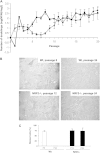
Figure 1.
Figure 1.
(A) Number of doublings per passage of wild-type (Wt) and Nrf2−/− murine embryonic fibroblasts (MEFs). Cells were cultured according to a 3T3 protocol. After each passage, cells were detached from culture plates and counted. Data are means ± SDs of measures taken at least in triplicate. A decline of Wt MEFs growth was observed between Passages 6 and 8. Cells resumed growth from Passage 9. Immortalization of Nrf2−/− was observed earlier (from Passage 5). (B) Senescence-associated (SA)-β-galactosidase staining. Wt cells harvested from passages with decreased cell growth were positive for SA-β-galactosidase, and the staining was lost in cells obtained from later passages, as an indicative of cellular immortalization. However, SA-β-galactosidase staining was still observed in immortalized Nrf2−/− MEFs. (C) Quantification of the number of cells positively stained for SA-β-galactosidase activity. About 150 cells in three different fields were counted. Differences between Wt and Nrf2−/− MEFs at P assage 24 were statistically different with p < .0001.
Figure 2.
Figure 2.
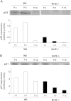
p53 and p21 levels. Cell extracts were prepared from wild-type (Wt) or Nrf2−/− murine embryonic fibroblasts (MEFs) obtained from the passages depicted in the figure and then probed for p53 (A) or p21 (B) by immunoblot. Results from a representative blot are shown for each case. Genetic deletion of Nrf2 resulted in the early loss of p53 and p21, the main hallmarks of MEFs immortalization.
Figure 3.
Figure 3.
Genetic deletion of Nrf2 decreases growth of immortalized murine embryonic fibroblasts (MEFs). The figure depicts the mean values of doublings number per passage. Data from all passages between 20 and 43 were used for calculations. Wild-type (Wt) MEFs, n = 142; Nrf2−/− MEFs, n = 94. Differences between Wt and Nrf2−/− MEFs were statistically different with p < .0001.
Figure 4.
Figure 4.
Cyclin E and A levels. Cell extracts were prepared from immortalized wild-type (Wt) or Nrf2−/− murine embryonic fibroblasts and then probed by immunoblot for cyclin levels. (A) Cyclin E. Both Wt and Nrf2−/− cells were obtained from Passage 27. (B) Cyclin A. Wt cells were obtained from Passage 29, whereas Nrf2−/− cells were obtained from Passage 28. For each antibody, the immunostained band was quantified with Quantity one software (Bio-Rad), and the results normalized to the density of the corresponding lane stained with Ponceau S. Results from representative blots are shown. Genetic deletion of Nrf2 resulted in a significant decrease of the levels of cyclin E but not of cyclin A.
Figure 5.
Figure 5.
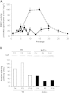
NQO1 and b5R levels. (A) Cytosolic fractions were obtained from wild-type (Wt) or Nrf2−/− murine embryonic fibroblasts (MEFs) obtained from the indicated passages and then assayed for NQO1 enzymatic activity. A significant increase of NQO1 activity was observed in Wt MEFs from Passages 10 to 15, at the beginning of cellular immortalization. On the other hand, as expected, NQO1 activity remained low in Nrf2−/− cells at any passage. Data are means ± SDs of measures taken in triplicate. (B) Membranous fractions were obtained from Wt or Nrf2−/− MEFs obtained from the indicated passages. Proteins were separated by sodium dodecyl sulfate–polyacrylamide gel electrophoresis and blotted onto nitrocellulose. Blots were then probed for b5R with a specific antiserum. The immunostained 34-kDa band was quantified with Quantity one software (Bio-Rad), and the results normalized to the density of the corresponding lane stained with Ponceau S. Results from a representative blot are shown.
Figure 6.
Figure 6.
Number of doublings per passage of wild-type (Wt) and Nrf2−/− murine embryonic fibroblasts in long-term cultures. Cells were cultured according to a 3T3 protocol. After each passage cells were detached from culture plates and counted. Data are means ± SDs of measures taken in triplicate. Genetic deletion of Nrf2 resulted in a significant shortening of cellular life span.
Figure 7.
Figure 7.
SirT1 levels. Cell extracts were prepared from murine embryonic fibroblasts obtained from the passages depicted in the figure and then probed for SirT1 by immunoblot. The immunostained band was quantified with Quantity one software (Bio-Rad), and the results normalized to the density of the corresponding lane stained with Ponceau S. Results from a representative blot are shown. Whereas SirT1 levels were higher in Nrf2−/− cells at passages less than 9, genetic deletion of Nrf2 resulted in a significant decrease of SirT1 levels in late passage immortalized cells.

Similar articles

Cited by

References

    1. Hayflick L. The limited in vitro lifetime of human diploid cell strains. Exp Cell Res. 1965;37:614–636. - PubMed
    1. Sherr CJ, DePinho RA. Cellular senescence: mitotic clock or culture shock? Cell. 2000;102:407–410. - PubMed
    1. Dirac AM, Bernards R. Reversal of senescence in mouse fibroblasts through lentiviral suppression of p53. J Biol Chem. 2003;278:11731–11734. - PubMed
    1. Todaro GJ, Green H. Quantitative studies of the growth of mouse embryo cells in culture and their development into established lines. J Cell Biol. 1963;17:299–313. - PMC - PubMed
    1. Kiyono T. Molecular mechanisms of cellular senescence and immortalization of human cells. Expert Opin Ther Targets. 2007;11:1623–1637. - PubMed

Publication types

Substances