. 2011 Jan;62(2):717-33.
doi: 10.1093/jxb/erq312. Epub 2010 Oct 25.

Water uptake by seminal and adventitious roots in relation to whole-plant water flow in barley (Hordeum vulgare L.)

Affiliations

Water uptake by seminal and adventitious roots in relation to whole-plant water flow in barley (Hordeum vulgare L.)

Thorsten Knipfer et al. J Exp Bot. 2011 Jan.

Abstract

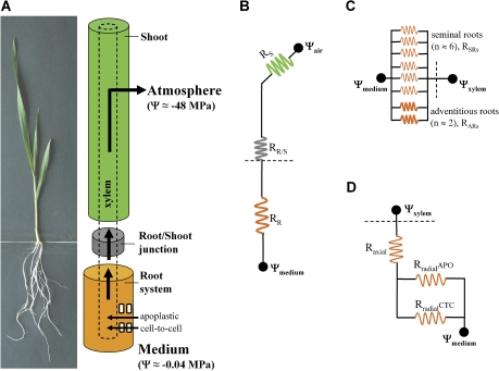
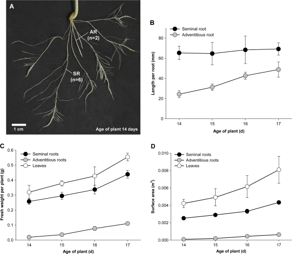
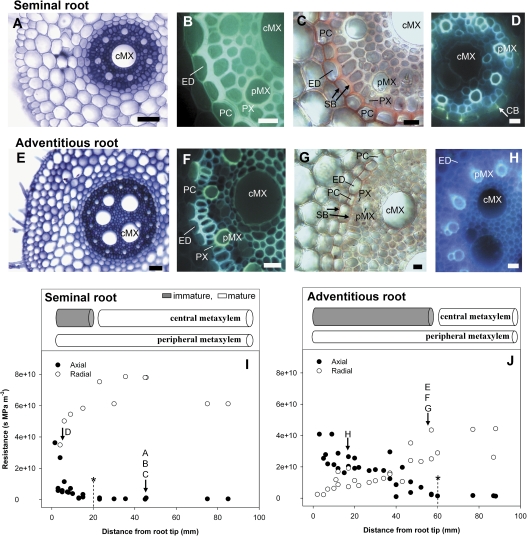
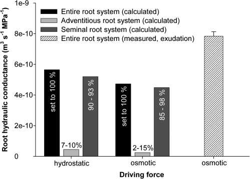
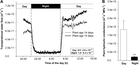
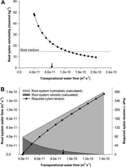
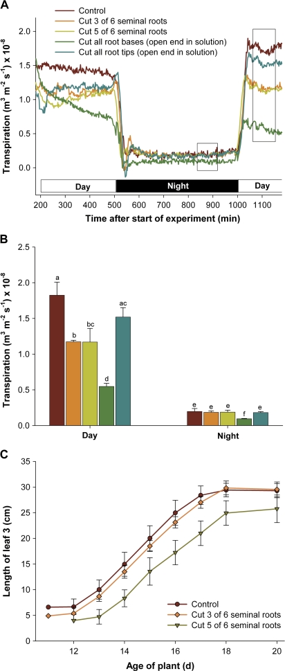
Prior to an assessment of the role of aquaporins in root water uptake, the main path of water movement in different types of root and driving forces during day and night need to be known. In the present study on hydroponically grown barley (Hordeum vulgare L.) the two main root types of 14- to 17-d-old plants were analysed for hydraulic conductivity in dependence of the main driving force (hydrostatic, osmotic). Seminal roots contributed 92% and adventitious roots 8% to plant water uptake. The lower contribution of adventitious compared with seminal roots was associated with a smaller surface area and number of roots per plant and a lower axial hydraulic conductance, and occurred despite a less-developed endodermis. The radial hydraulic conductivity of the two types of root was similar and depended little on the prevailing driving force, suggesting that water uptake occurred along a pathway that involved crossing of membrane(s). Exudation experiments showed that osmotic forces were sufficient to support night-time transpiration, yet transpiration experiments and cuticle permeance data questioned the significance of osmotic forces. During the day, 90% of water uptake was driven by a tension of about -0.15 MPa.


Figures

Fig. 1.
Fig. 1.
Hydraulic resistances along the soil–plant–atmosphere continuum. (A) The picture shows a 15-d-old barley plant as analysed in the present study. Water flow through the plant is driven by a difference in water potential, ΔΨ, between root medium (approximately –0.04 MPa) and atmosphere (approximately –48 MPa at 70% relative humidity and 21 °C). Radial water uptake into roots can occur along an apoplastic and a cell-to-cell path, the latter involving aquaporins. Water is transported axially along the xylem and may encounter resistances within the root system, at the root–shoot junction, or within the shoot (leaf). In the shoot, radial flow of water and exit into the atmosphere can be limited by the radial flow path or the conductance at the exit point (stomata, cuticle). (B–D) Major hydraulic resistances arranged analogously to an electrical circuit. (B) Resistance of the root system (RR), the root–shoot junction (RR/S), and the shoot (RS). (C) Within the root system, seminal and adventitious roots are treated as hydraulic resistances arranged in parallel (RSRs and RARs, respectively). (D) In each root, axial and radial hydraulic resistance are treated as being arranged in series (Raxial, Rradial); the radial resistance (Rradial) is divided into two resistances arranged in parallel, an apoplastic (RradialAPO), and a cell-to-cell resistance (RradialCTC).
Fig. 2.
Fig. 2.
Root and leaf growth in 14- to 17-d-old barley plants. (A) Typical root system showing (n=6) seminal roots (SR) and (n=2) adventitious roots (AR). (B) Average length of the main axis of individual seminal and adventitious roots. (C) Total fresh weight per plant of the entire set of seminal and adventitious roots and of leaves 1–3. (D) Total surface area per plant of the entire set of seminal and adventitious roots and of leaves 1–3. Results are means±SD (error bars) of values from (n=)12 plants, from three batches of plants. Where error bars seem to be absent, they are smaller than the symbol size. The surface area of roots was derived from an independently determined relationship between root fresh weight and surface area [seminal roots, n=17, fresh weight (g) ∼9.96×area (m2)+0.0054, r2=0.75; adventitious roots, n=7, fresh weight (g) ∼168.4×area (m2)+0.0033, r2=0.88]. (This figure is available in colour at JXB online.)
Fig. 3.
Fig. 3.
Anatomy and xylem development of seminal and adventitious roots of barley. (A–D) Cross-sections of seminal roots taken at 40–60 mm (A–C, lateral root zone) and 5–10 mm (D, tip region) from the tip. (A–C) Seminal roots have typically one central (cMX) and eight peripheral metaxylem (pMX) vessels, the latter located close to the much smaller protoxylem vessels (PX). The stelar region is highly lignified and metaxylem vessels are mature. The endodermis (ED) is mature and shows asymmetrically thickened cell walls, which is typical of state III of endodermis development (Enstone et al., 2003), in all cells including passage cells (PC). Wall depositions of suberin (SB, arrow) can be detected in all cells of the endodermis. (D) Close to the root tip only peripheral but not central metaxylem vessels are mature. Casparian bands can be detected (CB, arrow). (E–H) Cross-sections of adventitious roots taken at 40–60 mm (E–G, root hair region) and 15–20 mm (H, tip region) from the root tip. (E–G) Adventitious roots have typically 5–7 central and 14 peripheral metaxylem vessels. The stelar region shows some lignification. The endodermis shows secondary wall thickening except in passage cells. There are fewer suberin depositions in adventitious compared with seminal roots and depositions are lacking from some passage cells. Central metaxylem appears less mature than in seminal roots. (H) Closer to the root tip, only peripheral but not central metaxylem vessels appear mature. Sections shown in (A) and (E) were stained with Toluidine Blue and viewed under bright light; sections in (B) and (F) were stained with berberine hemisulfate and counterstained with Toluidine Blue and viewed under fluorescence light (390–420 nm) to visualize Casparian bands and xylem development (Brundrett et al., 1988). Sections in (C) and (G) were stained with Sudan Red 7B and viewed under bright light to visualize depositions of suberin (Brundrett et al., 1991). (I–J) Root pressure probe analyses of axial and radial hydraulic resistance (inverse of conductance) along (I) seminal and (J) adventitious roots. The axial hydraulic resistance was experimentally determined from half-times obtained through root pressure probe experiments where roots were cut back successively and in between measurements (data points) from the tip (see Frensch and Steudle, 1989). The radial resistance was calculated as the difference between the overall root resistance and the axial resistance for a particular location. (I) In seminal roots, the axial resistance decreases to very low values beyond 20 mm from the tip, whereas the radial resistance increases. This shows that central metaxylem vessels become fully mature and the endodermis fully developed at ∼20 mm (as indicated by asterisk). (J) In adventitious roots, changes in axial and radial resistance (and corresponding changes in metaxylem and endodermis development) occur up to 60 mm from the tip (asterisk). Results are pooled from three root analyses each, and the location of cross-sections shown in A–H is indicated. Scale bars: (A) 55 μm, (B–D) 15 μm, (E) 75 μm, and (F–H) 25 μm.
Fig. 4.
Fig. 4.
Contribution of seminal and adventitious roots to water uptake in 14- to 17-d-old barley plants in dependence on the driving force (osmotic, hydrostatic). Hydraulic conductance was determined through osmotic and hydrostatic experiments for individual seminal and adventitious roots (see Table 3). The average values of these experiments were then used to calculate the hydraulic conductance of a typical seminal and a typical adventitious root system of a barley plant, containing 6–7 (average 6.5) seminal and 2–4 (average 3) adventitious roots, respectively. The sum of the two gave the conductance of the entire root system of a plant. Percentage figures give the contribution of the conductance of the seminal and adventitious root system to the conductance of the entire root system of a barley plant. The range of conductance values, as calculated from the range of means given in Table 3 was as follows (unit: m3 s−1 MPa−1×10⁻¹⁰): hydrostatic force, seminal roots, 4.2–6.1; adventitious roots, 0.45 (results from only one analytical method used); entire root system of plant, 4.7–6.6; osmotic force, seminal roots, 1.8–7.7; adventitious roots, 0.18–0.30; entire root system of plant, 2.0–8.0. Also shown is an experimentally (exudation) determined osmotic hydraulic conductance for an entire barley root system; average and SD (error bars) of four independent root analyses.
Fig. 5.
Fig. 5.
Day- and night-time transpiration, and whole-plant hydraulic conductance of hydroponically grown barley plants. (A) Typical trace of gravimetrically determined transpiration of two barley plants (14 d and 15 d old at the start of measurement). Average (± SD) day and night transpiration rates of four plants are shown in the insert. (B) Whole-plant hydraulic conductance during the day and night; means±SD of four plants (***, P<0.001, Student's t-test).
Fig. 6.
Fig. 6.
Calculation of osmotic and hydrostatic forces required to drive root water uptake during the day in transpiring barley plants. (A) Osmolality of exudate of seminal roots was determined as part of exudation experiments. Since the exudate flow rate was ∼40% of the rate of transpirational water loss, and since xylem solute concentrations decrease with increasing flow rate (e.g. Munns and Passioura, 1984), a simulation was carried out in which xylem osmolality was calculated in dependence of flow rate (for details, see Equation 13, Supplementary File S1). (B) Using the relationship shown in (A) a water flow driven through osmotic forces (Root system osmotic) was calculated in dependence of transpirational water flow. The difference between transpirational water flow and osmotically driven flow is water flow driven by a tension (Root system hydrostatic). Based on the linear relationship between hydrostatic flow rate and applied tension, as previously determined through vacuum perfusion experiments (Knipfer and Fricke, 2010), this allowed us to calculate the tension required to drive daytime water uptake—in addition to water uptake driven through osmotic gradients. For a transpirational water flow of 8.8×10⁻¹¹ m3 s−1 as measured in the growth chamber for barley plants during the day (see arrow), osmotic forces drove 10% while hydrostatic forces (tension of about –150 kPa) drove the remaining 90% of root water uptake.
Fig. 7.
Fig. 7.
Day- and night-time transpiration, and leaf growth in hydroponically grown barley plants with (partially) excised root system. Barley plants had an intact seminal root system (control); or had three or five of six seminal roots removed close to the root base, just above the nutrient solution (the cut was sealed with Vaseline); or all six roots removed close to the base, with the cut end extending ∼2 cm into the nutrient solution; or had the tip 2 cm of all six seminal roots removed, with the cut end extending into the nutrient solution. Only seminal roots were manipulated, since adventitious roots were developed little and contributed little to water uptake in control plants (compare with Fig. 4). (A) Continuous recordings of transpiration. Transpirational water loss was related to total leaf area as determined at the end of experiments. The part of traces that is boxed in was used to calculate average transpiration rates during the day and night shown in (B) [means±SD of five (control) and three (treatments) plant analyses]. (C) Growth of leaf 3 in plants with an intact root system (control) and in plants that had three or five of six seminal roots excised; means±SD of 12 plant analyses from three batches of plants. In (B) statistically significant differences between transpiration rates are indicated by different letters (P<0.05, Student's t-test).

Similar articles

Cited by

References

    1. Bramley H, Turner NC, Turner DW, Tyerman SD. Roles of morphology, anatomy, and aquaporins in determining contrasting hydraulic behavior of roots. Plant Physiology. 2009;150:348–364. - PMC - PubMed
    1. Brundrett MC, Enstone DE, Peterson CA. A berberine-aniline blue staining procedure for suberin, lignin, and callose in plant tissue. Protoplasma. 1988;146:133–142.
    1. Brundrett MC, Kendrick B, Peterson CA. Efficient lipid staining in plant material with Sudan red 7B or fluorol yellow 088 in polyethylene glycol-glycerol. Biotechnology and Histochemistry. 1991;66:111–116. - PubMed
    1. Caird MA, Richards JH, Donovan LA. Nighttime stomatal conductance and transpiration in C3 and C4 plants. Plant Physiology. 2007;143:4–10. - PMC - PubMed
    1. Ehlert C, Maurel C, Tardieu F, Simonneau T. Aquaporin-mediated reduction in maize root hydraulic conductivity impacts cell turgor and leaf elongation even without changing transpiration. Plant Physiology. 2009;150:1093–1104. - PMC - PubMed

Publication types