. 2010 Dec;16(12):2464-73.
doi: 10.1261/rna.2346510. Epub 2010 Oct 25.

Short RNA duplexes guide sequence-dependent cleavage by human Dicer

Affiliations

Short RNA duplexes guide sequence-dependent cleavage by human Dicer

Lucien Bergeron Jr et al. RNA. 2010 Dec.

Abstract

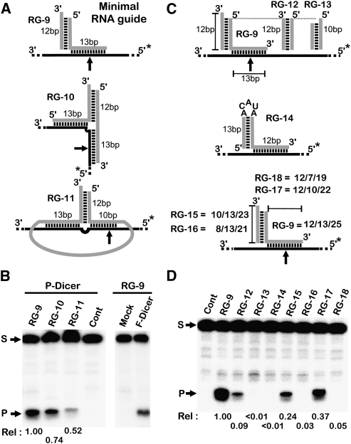
Dicer is a member of the double-stranded (ds) RNA-specific ribonuclease III (RNase III) family that is required for RNA processing and degradation. Like most members of the RNase III family, Dicer possesses a dsRNA binding domain and cleaves long RNA duplexes in vitro. In this study, Dicer substrate selectivity was examined using bipartite substrates. These experiments revealed that an RNA helix possessing a 2-nucleotide (nt) 3'-overhang may bind and direct sequence-specific Dicer-mediated cleavage in trans at a fixed distance from the 3'-end overhang. Chemical modifications of the substrate indicate that the presence of the ribose 2'-hydroxyl group is not required for Dicer binding, but some located near the scissile bonds are needed for RNA cleavage. This suggests a flexible mechanism for substrate selectivity that recognizes the overall shape of an RNA helix. Examination of the structure of natural pre-microRNAs (pre-miRNAs) suggests that they may form bipartite substrates with complementary mRNA sequences, and thus induce seed-independent Dicer cleavage. Indeed, in vitro, natural pre-miRNA directed sequence-specific Dicer-mediated cleavage in trans by supporting the formation of a substrate mimic.


Figures

FIGURE 1.
FIGURE 1.
Reconstitution of Dicer substrates in trans using short single-stranded RNAs. (A) Schematic representation of the short single-stranded RNA guides (RG, in gray) used to target the Dicer cleavage in B. The positions of the observed cleavage sites are indicated by the arrows along the target (black line). The nonproductive oligoribonucleotide is marked with an X. (*) The position of the 5′-32P. (B) Different single-stranded RNAs (RG1-3) were incubated with either 5′-end-labeled 151-nt-long single-stranded RNA (LS) or short 70-nt single-stranded RNA (SS) (both derived from the Caspase 8 mRNA) in either the presence (RG-X) or absence (Cont) of specific guides. The positions of the input substrates (LS or SS), as well as of the cleavage products (P1-3), are indicated. The percent cleavage for each reaction is indicated at the bottom. (C) Formation of perfect RNA duplexes is not required for Dicer cleavage. Substrates carrying insertions of 4–15 nt upstream of the predicted cleavage sites were generated and tested for cleavage in vitro (D). (Gray boxes) The positions of the predicted cleavage sites; (arrows) the observed cleavage sites. (S) Input substrates; [P(4-8)] the products. The cleavage velocities relative (Rel) to that of the unmutated substrate (RG-4) are shown at the bottom. All experiments are repeated at least three times with a maximum error of the rate of +0.05.
FIGURE 2.
FIGURE 2.
Directing Dicer cleavage using a fixed binding site. (A) Schematic representation of the different RNAs (in gray) used to guide Dicer cleavage in trans. An RNA stem with a fixed sequence terminating in a 3′-overhang was fused to single-stranded RNA fragments capable of forming either a short RNA duplex (RG-9), a long RNA duplex (RG-10), or a long RNA duplex with joined termini (RG-11) with a common target sequence. (*) The position of the 5′-32P. (B) The different RNA guides illustrated in A were incubated with 5′-end-labeled target RNA (black line) derived from the Caspase 8 mRNA and partially purified commercial Dicer protein (P-Dicer) in either the absence (Cont) or the presence of the corresponding guide. Cleavage of the RNA substrates was also performed using immunoprecipitated Flag-tagged Dicer (F-Dicer), and a mock precipitation was included as a control (i.e., RG-9 was presented in both reactions). (C) Schematic representation of the different RNA molecules designed to characterize the features of the RNA stem required to guide Dicer cleavage in trans. (D) The RNA target complexes with guide RNAs (from C) were incubated with partially purified Dicer as described in B. (S) Position of the substrate; (P) position of the cleavage product. The relative velocities (Rel) are indicated at the bottom. All experiments were repeated at least three times with a maximum error of the rate of +0.05.
FIGURE 3.
FIGURE 3.
Effect on reactivity of 2′-OMe groups into the primary Dicer binding site. (A) The impact of the 2′-OH groups on substrate selection and cleavage by Dicer was examined. Different RNA oligonucleotides were synthesized with 2′-OMe substitutions in the Dicer binding site (RG-19), within the guide sequence (RG-20), or in both binding and guide sequences (RG-21). (Green lines) RNA sequences present in the RG; (blue lines) the sequences containing the 2′-OMe groups; (*) the position of the 5′-32P. (B) Cleavage reactions were conducted as described in Figure 2 using 5′-end-labeled substrates as targets (black line). (Rel) The cleavage velocities relative to that obtained with RG-9. The values shown represent the average of at least two experiments with a maximum error of the rate of +0.05. (S) Position of the substrate; (P) positions of the products. (C) Summary of the cleavage activities obtained with various RNA guides carrying either single or multiple 2′-OMe groups near the Dicer cleavage site. (Red) The position of the 2′-OMe group; (arrowheads) the positions of Dicer cleavage; (black arrows) strong cleavage; (open arrows) medium cleavage; (gray arrows) weak cleavage. For clarity, the fixed binding sites were drawn only for the first three examples.
FIGURE 4.
FIGURE 4.
Analysis of Dicer-dependent RNA-guided cleavage. (A) The presence of a single base mismatch on the binding arm of the guide abolishes Dicer cleavage. Various Dicer RNA guides (in gray) carrying mutations that disrupt base-pairing in either Dicer binding site, or in the targeting sequence (left panel), were synthesized and tested for cleavage using a 5′-end-labeled target RNA as described in Figure 2 (right panels). (*) The position of the 5′-32P. (S) The positions of the input substrates; (P) the positions of the cleavage products. (B) Short duplex RNAs direct precise site-specific cleavage. RNA guides (in gray) targeting different positions in a single target RNA (black line, left panel) were synthesized and tested for cleavage (right panel) as described in A. The target RNA was incubated in the presence of partially purified Dicer and the strand guide harboring the binding arm in either the presence (+) or the absence (−) of the complementary Univ strand forming the 3′-overhang stem. For the control condition (Cont), the RG is missing, and for the Univ condition, the binding arm strand was omitted. The positions and the names of the guide RNAs driving the cleavage are shown on the left of the gel. (Rel) The cleavage velocities relative to that obtained with RG-9, represents the average of at least two experiments with a maximum error of the rate of +0.05.
FIGURE 5.
FIGURE 5.
A micro-RNA-like structure guides Dicer cleavage in vitro. (A) Representations of the different designs of the RNA guides produced in order to test whether or not an miRNA-like structure may drive Dicer cleavage in vitro. The miRNA-like guide RNAs are shown either in the absence (on top), or the presence (at bottom), of the target RNA (black line). B1 (blue), B2 (yellow), and K (red) indicate, respectively, the binding arm or the equivalent of the seed sequence in an miRNA, the strand complementary to the target site, and the sequence pairing with the binding arm. RG-34 mimics the structure of a mature miRNA, while RG-35 represents a short version of a pre-miRNA. (*) The position of the 5′-32P. (B) RNA cleavage using an miRNA-like construct as guide. The miRNA-like guide RNAs, which contain either the good corresponding sequences (+) or unrelated sequences (−), were incubated with the 5′-end-labeled RNA target. (Rel) The cleavage velocities relative to that obtained with RG-9, represents the average of at least two experiments with a maximum error of the rate of +0.05.
FIGURE 6.
FIGURE 6.
Seed-independent pairing of pre-miRNA with mRNA induces Dicer dependent RNA degradation. (A) Schematic representation of the natural processing pathway of the hsa-pre-miR-500 (upper part), and a possible alternative target mRNA-independent degradation pathway (lower part). Oligoribonucleotides carrying the natural hsa-pre-miR-500 sequence were synthesized with 3 nt near the 3′-end mutated in order to allow for run-off transcription (blue circles). The blue and orange strands correspond to the target binding arms. In the left panel, the sites of Dicer cleavage are indicated by arrows (sites 1 and 2 produce P1 and P2 in B). (Black line) The RNA target. (*) The position of the 5′-32P. (B) Dicer accurately processes hsa-pre-mir-500 in vitro. 5′-End labeled hsa-pre-mir-500 (pm) was incubated in either the absence (−) or the presence (+) of partially purified Dicer (P-Dicer), or was incubated with immunoprecipitated Flag-tagged Dicer (F-Dicer). The cleavage reaction was also performed in the presence of a 10× excess of the target sequence complementary to the binding arms of the hsa-pre-mir-500 (10×). The pre-miRNA cleavage products (P1 and P2) are indicated. (C) Pre-miRNA targets Dicer cleavage to long RNA fragments in vitro. The hsa-pre-mir-500 was incubated with a 5′-end-labeled 151-nt-long RNA (S) carrying a sequence complementary to either the binding arms of the miRNA (Target, lane 1) or an unrelated sequence (US, lane 4). (P) The target product. In lane 2, the target and the pre-miRNA were both labeled. The mature miRNA produced by Dicer cleavage was incubated with the long RNA target in lane 3. The results shown are representative of at least two independent experiments. (Open triangle) An unidentified cleavage product.

Similar articles

Cited by

References

    1. Ahmed F, Ansari HR, Raghava GP 2009. Prediction of guide strand of microRNAs from its sequence and secondary structure. BMC Bioinformatics 10: 105 doi: 10.1186/1471-2105-10-105 - PMC - PubMed
    1. Berezikov E, Chung WJ, Willis J, Cuppen E, Lai EC 2007. Mammalian mirtron genes. Mol Cell 28: 328–336 - PMC - PubMed
    1. Calin-Jageman I, Amarasinghe AK, Nicholson AW 2001. Ethidium-dependent uncoupling of substrate binding and cleavage by Escherichia coli ribonuclease III. Nucleic Acids Res 29: 1915–1925 - PMC - PubMed
    1. Carbonell A, Martinez de Alba AE, Flores R, Gago S 2008. Double-stranded RNA interferes in a sequence-specific manner with the infection of representative members of the two viroid families. Virology 371: 44–53 - PubMed
    1. Carmell MA, Hannon GJ 2004. RNase III enzymes and the initiation of gene silencing. Nat Struct Mol Biol 11: 214–218 - PubMed

Publication types