. 2010 Nov 1;191(3):505-21.
doi: 10.1083/jcb.201007098. Epub 2010 Oct 25.

Pom121 links two essential subcomplexes of the nuclear pore complex core to the membrane

Affiliations

Pom121 links two essential subcomplexes of the nuclear pore complex core to the membrane

Jana M Mitchell et al. J Cell Biol. .

Abstract

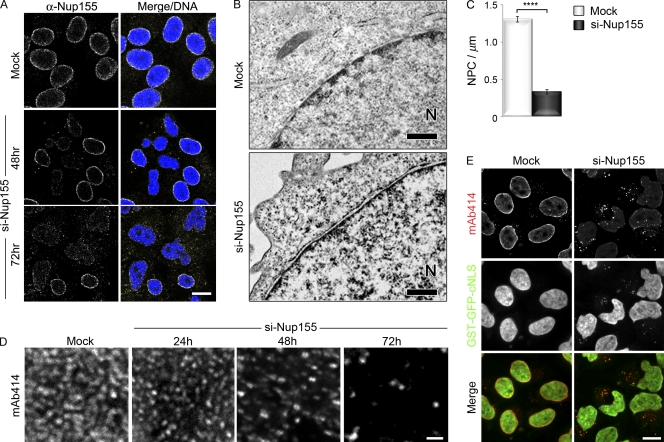
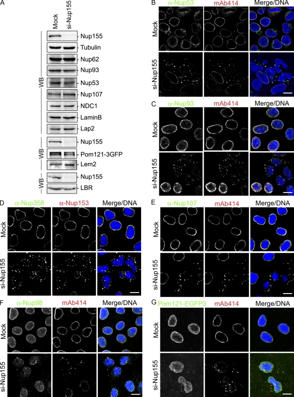
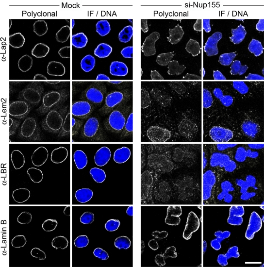
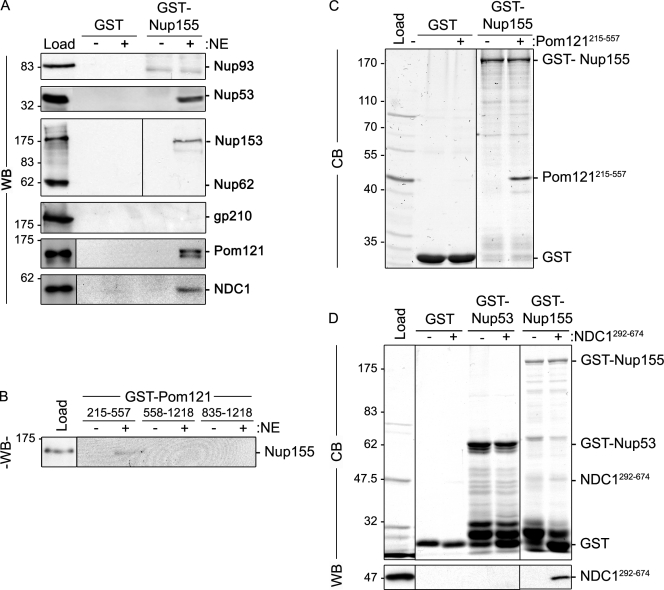
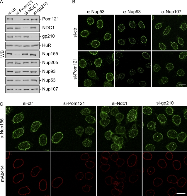
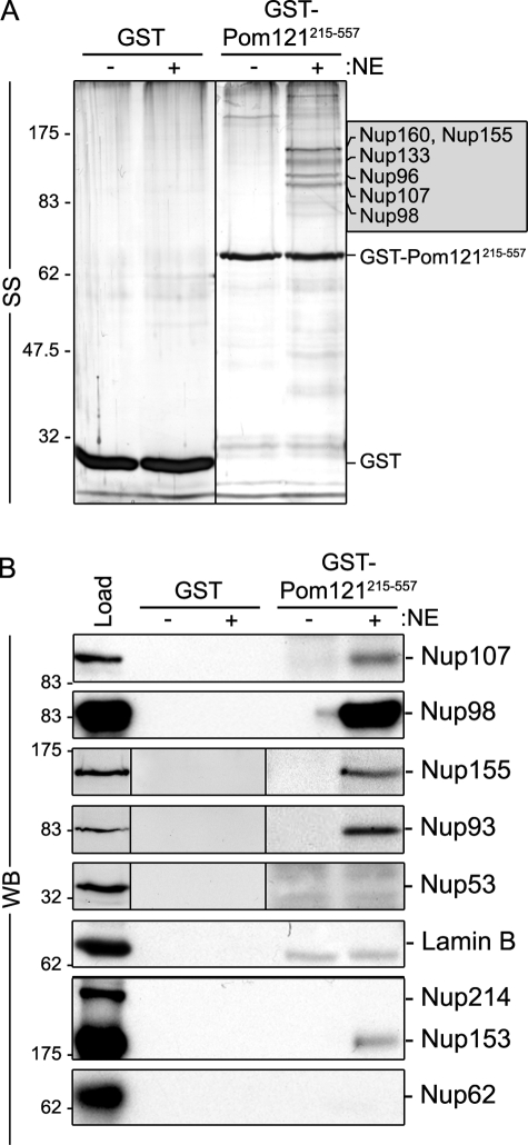
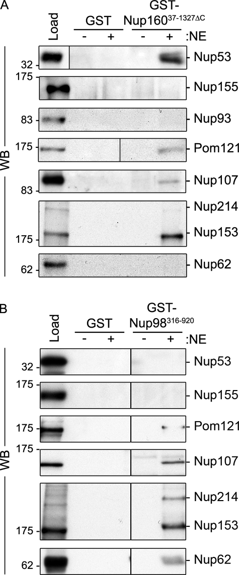
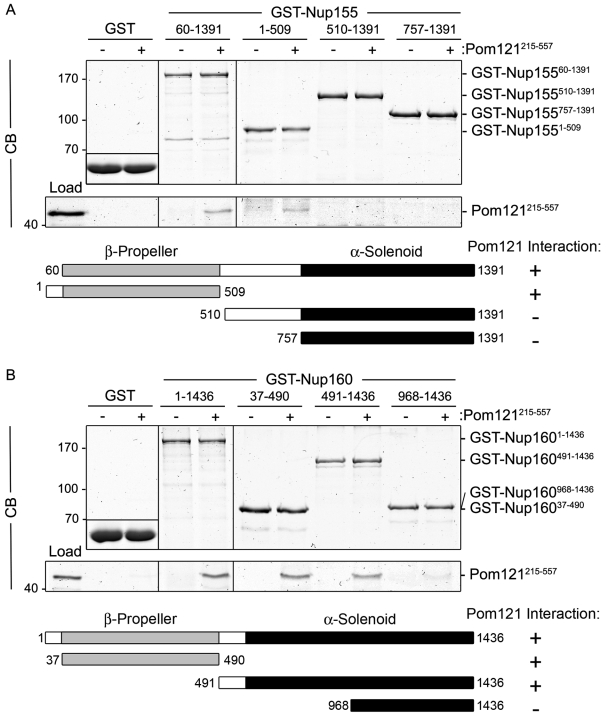
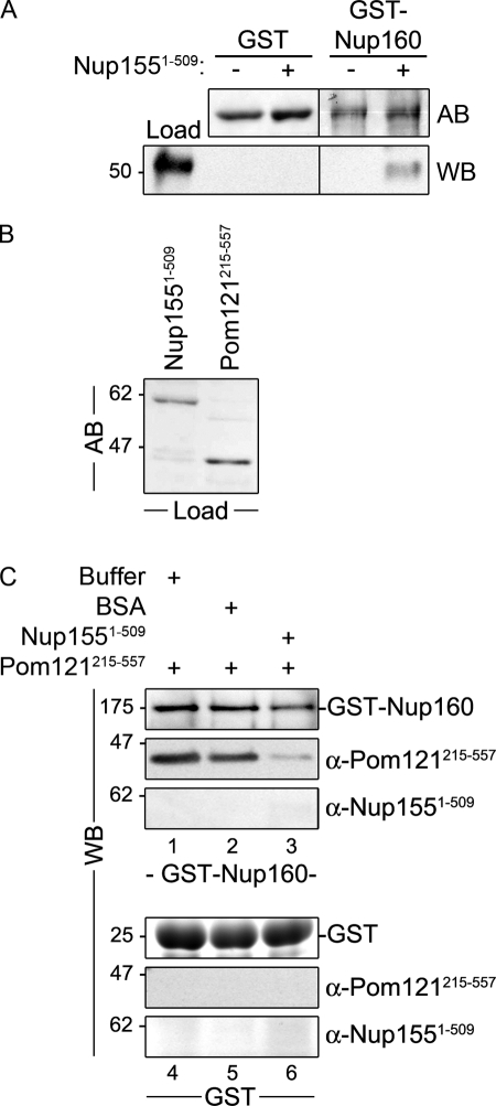
Nuclear pore complexes (NPCs) control the movement of molecules across the nuclear envelope (NE). We investigated the molecular interactions that exist at the interface between the NPC scaffold and the pore membrane. We show that key players mediating these interactions in mammalian cells are the nucleoporins Nup155 and Nup160. Nup155 depletion massively alters NE structure, causing a dramatic decrease in NPC numbers and the improper targeting of membrane proteins to the inner nuclear membrane. The role of Nup155 in assembly is likely closely linked to events at the membrane as we show that Nup155 interacts with pore membrane proteins Pom121 and NDC1. Furthermore, we demonstrate that the N terminus of Pom121 directly binds the β-propeller regions of Nup155 and Nup160. We propose a model in which the interactions of Pom121 with Nup155 and Nup160 are predicted to assist in the formation of the nuclear pore and the anchoring of the NPC to the pore membrane.


Figures

Figure 1.
Figure 1.
Depletion of Nup155 alters nuclear morphology and decreases NPC number. HeLa cells were treated with transfection reagent alone (Mock) or the Nup155-specific siRNA-targeting exon 27 (si-Nup155) as indicated. (A) Nup155 levels were assayed by indirect immunofluorescence using anti-Nup155 antibodies at the indicated times and DNA visualized with DRAQ5 (Merge/DNA). Bar, 10 µm. (B and C) Mock- or Nup155-depleted HeLa cells were processed for transmission electron microscopy 72 h after transfection. The position of the nucleoplasm is indicated (N). Bar, 0.5 µm. (C) The number of NPCs per micron of NE was determined by examining individual nuclear sections (n > 48 sections per condition). The average number of NPCs per micron of NE is shown. The P value (P < 0.0001, represented by asterisks) was calculated using a Welch corrected unpaired t test. Error bars indicate standard error. (D) NPCs at the nuclear surface of Nup155-depleted cells were visualized by indirect immunofluorescence using mAb414 at the indicated times after siRNA treatment. Bar, 1 µm. (E) HeLa cells expressing GST-GFP-cNLS were incubated in the presence of transfection reagent alone (Mock) or with Nup155-specific siRNAs. 72 h after transfection, GST-GFP-cNLS was visualized together with mAb414-reactive nups detected by immunofluorescence. Bar, 10 µm.
Figure 2.
Figure 2.
Depletion of Nup155 in HeLa cells leads to the mislocalization of nucleoporins. HeLa cells were incubated in the presence of transfection reagent alone (Mock) or Nup155-specific siRNA (si-Nup155) for 72 h. (A) Total cell extracts from Mock- and Nup155-depleted cells were assayed by Western blot (WB) analysis using antibodies directed against the indicated proteins. Note: the bottom blot panels are from separate experiments. (B–F) The cellular distribution of various nups was visualized by indirect immunofluorescence using the indicated polyclonal anti-nup antibodies and the mouse monoclonal mAb414 (mAb414 detects Nup62, Nup153, Nup214, and Nup358) or mouse monoclonal anti-Nup153. (G) In Mock- or Nup155-specific siRNA-transfected cells, Pom121-EGFP3 was visualized directly and compared with mAb414-reactive nups detected by immunofluorescence. In all panels merged fluorescence images and DNA visualized with DRAQ5 are shown (Merge/DNA). Bar, 10 µm.
Figure 3.
Figure 3.
Nup155 depletion alters targeting of INM proteins. HeLa cells were incubated in the presence of transfection reagent alone (Mock) or Nup155-specific siRNA (si-Nup155) for 72 h and then processed for immunofluorescence microscopy. The indicated INM proteins and lamin B were visualized using specific polyclonal antibodies and DNA was visualized using DRAQ5 (Merge/DNA). Bar, 10 µm.
Figure 4.
Figure 4.
Identification of Nup155-interacting proteins. (A) GST-Nup155 or GST alone were bound to glutathione–Sepharose beads and then incubated in the presence (+) or absence (−) of solubilized rat liver NE extracts. Bound proteins were eluted from beads using SDS-sample buffer. Interacting nups were detected by Western blot (WB) using antibodies directed against the indicated proteins. mAb414 was used to detect Nup62 and Nup153. Approximately 5% of the NE extract loaded on each column was resolved in the lane marked Load. (B) GST-tagged truncations of the pore-facing domain of Pom121 (amino acid residues 215–557, 558–1218, or 835–1218) were incubated with (+) or without (−) rat liver NE extracts. Interacting proteins were eluted with SDS-sample buffer. Polypeptides were resolved by SDS-PAGE and analyzed by Western blotting (WB) using a Nup155-specific polyclonal antibody. Approximately 5% of the NE extract loaded on each column was resolved in the lane marked Load. (C and D) Glutathione–Sepharose beads with attached GST-Nup155, GST-Nup53, or GST alone were incubated with (+) or without (−) purified recombinant Pom121215–557 (C) or Ndc1292–674 (D). Bound proteins were processed as described in A and polypeptides were detected using Coomassie blue staining (CB) or Western blot (WB) using anti-NDC1 antibodies. The lanes marked Load contain ∼50% of the total purified Pom121215–557 (C) or NDC1292–674 (D) loaded on the beads. To the right of each panel, the point at which the named recombinant protein migrates in the appropriate lane is indicated. Mass markers are in kilodaltons.
Figure 5.
Figure 5.
Depletion of Pom121 reduces cellular levels of Nup155. HeLa cells were incubated in the presence of control siRNAs (si-ctr) or siRNAs targeting Pom121, NDC1, or gp210 as indicated. (A) Cells were harvested and analyzed by Western blotting (WB) using antibodies directed against the indicated proteins. α-HuR was used as a loading control. (B) HeLa cells were processed for immunofluorescence microscopy using anti-Nup53, anti-Nup93, or anti-Nup107 polyclonal antibodies. Bar, 10 µm. (C) Cells were processed for immunofluorescence microscopy and interrogated using anti-Nup155 or mAb414 antibodies. Bar, 10 µm.
Figure 6.
Figure 6.
Pom121 interacts with Nup155 and the Nup107–160 complex in vitro. Bead-bound GST-Pom121215–557 or GST alone was incubated with (+) or without (−) rat liver NE extracts. Interacting proteins were eluted with SDS-sample buffer. Polypeptides were resolved by SDS-PAGE and visualized by silver staining (SS; A) or analyzed by Western blotting (WB; B) using antibodies directed against the indicated proteins. mAb414 was used to detect Nup62, Nup153, and Nup214. Approximately 5% of the NE extract loaded on each column was resolved in the lane marked Load. Prominent bands were analyzed by mass spectrometry (see Fig. S3) and the predominant species identified in the 90–160-kD range are indicated in the shaded box. Molecular mass markers are indicated in kilodaltons.
Figure 7.
Figure 7.
Identification of nups that bind Nup160 and Nup98. GST-Nup16037–1327ΔC (A), GST-Nup98316–920 (B), or GST alone were bound to glutathione–Sepharose beads and incubated in the presence (+) or absence (−) of solubilized rat liver NE extracts. Bound proteins were eluted from beads using SDS-sample buffer. Interacting nups were detected by Western blot (WB) using antibodies directed against the indicated proteins. mAb414 was used to detect Nup62, Nup153, and Nup214. Approximately 5% of the NE extract loaded on each column was resolved in the lane marked Load. Molecular mass markers are indicated in kilodaltons.
Figure 8.
Figure 8.
Pom121 interacts directly with Nup98 and N-terminal domains of Nup155 and Nup160. Purified Pom121215–557 was incubated with bead-bound (A) GST-Nup15560–1391, GST-Nup1551–509, GST-Nup155510–1391, and GST-Nup155757–1391, or (B) GST-Nup1601–1436, GST-Nup16037–490, GST-Nup160491–1436, and GST-Nup160968–1436. Shown in each panel are controls where Pom121215–557 was incubated with GST alone (shown as inset). To obtain the results shown in each panel, bead-bound proteins were eluted using SDS-sample buffer, resolved by SDS-PAGE, and visualized by Coomassie blue (CB) staining. The lane marked Load contains ∼10% of the total purified Pom121215–557 loaded on the beads. To the right of each panel, the point at which the named protein migrates in the appropriate lane is indicated. Schematic representations of Nup155 (A) and Nup160 (B) fragments and their observed binding to Pom121215–557 are summarized. Mass markers are in kilodaltons.
Figure 9.
Figure 9.
Binding of Nup155 to Pom121 prevents their interactions with Nup160. (A) Bead-bound GST-Nup160 or GST alone was incubated with purified Nup1551–509. Bound proteins were eluted using SDS-sample buffer and resolved by SDS PAGE. Levels of GST and GST-Nup160 were visualized using amido black (AB). Nup1551–509 was detected using a Nup155-specific polyclonal antibody. The lane marked Load contains ∼6% of the total purified Nup1551–509 loaded on the beads. (B and C) Purified Pom121215–557 and Nup1551–509 (∼25% of the total Load protein is shown in B after amido black [AB] staining) were mixed and allowed to interact for 30 min before addition to bead-bound GST-Nup160 or GST alone (lanes 3 and 6). Note: to perform the binding reactions under similar conditions, Pom121215–557 was supplemented with BSA to a total protein concentration similar to that of combined Pom121215–557–Nup1551–509. Bound proteins were eluted using SDS-sample buffer and resolved by SDS-PAGE. Western blotting (WB) using anti-Pom121, anti-Nup155, or anti-GST-Nup160 antibodies was used to detect the presence of each protein. Mass markers are indicated in kilodaltons.

Similar articles

Cited by

References

    1. Aaronson R.P., Blobel G.. 1974. On the attachment of the nuclear pore complex. J. Cell Biol. 62:746–754. 10.1083/jcb.62.3.746 - DOI - PMC - PubMed
    1. Abramoff M.D., Magelhaes P.J., Ram S.J.. 2004. Image processing with ImageJ. Biophotonics International. 11:36–42.
    1. Akey C.W. 1989. Interactions and structure of the nuclear pore complex revealed by cryo-electron microscopy. J. Cell Biol. 109:955–970. 10.1083/jcb.109.3.955 - DOI - PMC - PubMed
    1. Akey C.W., Radermacher M.. 1993. Architecture of the Xenopus nuclear pore complex revealed by three-dimensional cryo-electron microscopy. J. Cell Biol. 122:1–19. 10.1083/jcb.122.1.1 - DOI - PMC - PubMed
    1. Alber F., Dokudovskaya S., Veenhoff L.M., Zhang W., Kipper J., Devos D., Suprapto A., Karni-Schmidt O., Williams R., Chait B.T., et al. . 2007a. Determining the architectures of macromolecular assemblies. Nature. 450:683–694. 10.1038/nature06404 - DOI - PubMed

Publication types

MeSH terms