. 2010 Nov 1;191(3):677-91.
doi: 10.1083/jcb.201006052. Epub 2010 Oct 25.

Neutrophil elastase and myeloperoxidase regulate the formation of neutrophil extracellular traps

Affiliations

Neutrophil elastase and myeloperoxidase regulate the formation of neutrophil extracellular traps

Venizelos Papayannopoulos et al. J Cell Biol. .

Abstract

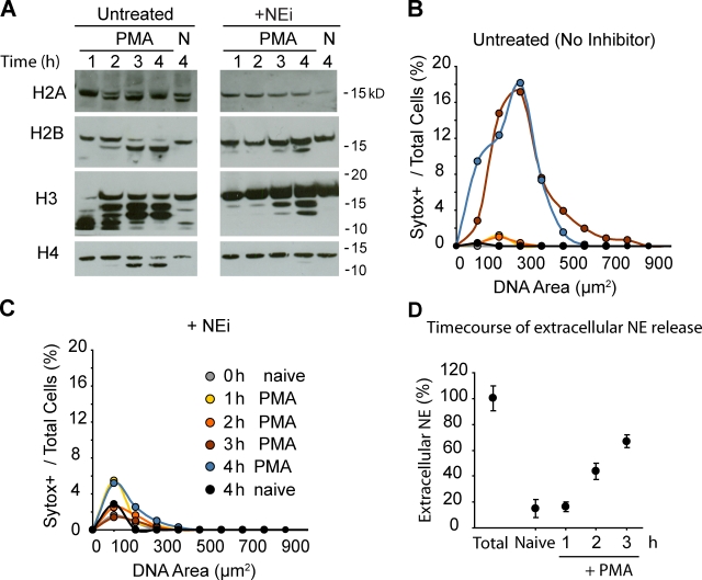
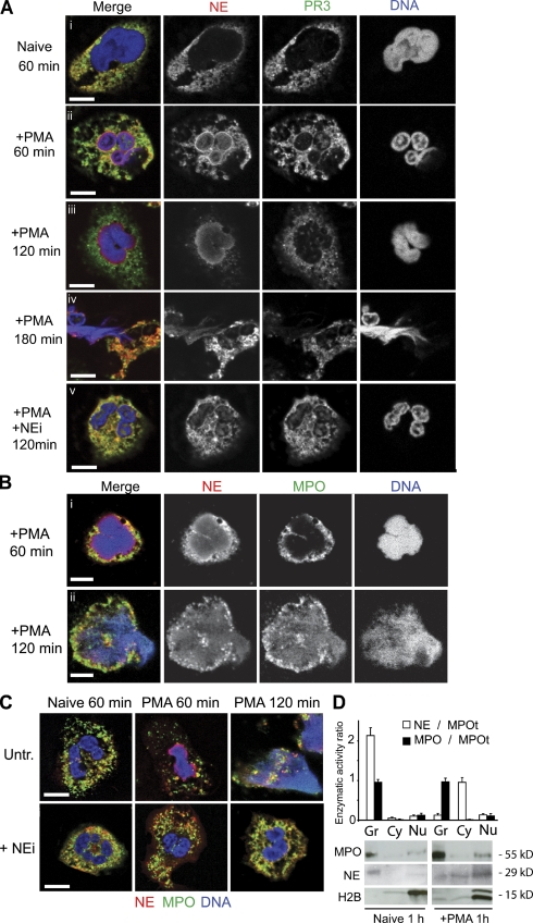
Neutrophils release decondensed chromatin termed neutrophil extracellular traps (NETs) to trap and kill pathogens extracellularly. Reactive oxygen species are required to initiate NET formation but the downstream molecular mechanism is unknown. We show that upon activation, neutrophil elastase (NE) escapes from azurophilic granules and translocates to the nucleus, where it partially degrades specific histones, promoting chromatin decondensation. Subsequently, myeloperoxidase synergizes with NE in driving chromatin decondensation independent of its enzymatic activity. Accordingly, NE knockout mice do not form NETs in a pulmonary model of Klebsiella pneumoniae infection, which suggests that this defect may contribute to the immune deficiency of these mice. This mechanism provides for a novel function for serine proteases and highly charged granular proteins in the regulation of chromatin density, and reveals that the oxidative burst induces a selective release of granular proteins into the cytoplasm through an unknown mechanism.


Figures

Figure 1.
Figure 1.
NE cleaves histones and promotes nuclear decondensation in vitro. (A) Nuclei isolated from neutrophils were incubated in buffer or in neutrophil-derived LSS lysates for 120 min at 37°C and labeled with Sytox green. Bar, 10 µm. (B) Neutrophil extracts are sufficient to decondense nuclei from other cell types. Nuclear decondensation of LSS extracts from HL-60 cells differentiated with RA, HeLa cells, PBMCs, and neutrophils were tested with nuclei isolated from neutrophils (Neut). The nuclear area was quantified using ImageJ image processing software. Circles denote the median area and the bars indicate the range of the nuclear area values, calculated from the standard deviation of each dataset. (C) Decondensation activity is stored in azurophilic granules. Decondensation of nuclei treated with buffer, neutrophil LSS, neutrophil HSS, HSP, and the granular subfractions of gelatinase (1), specific (2), and azurophilic (3) granules were purified over a discontinuous Percoll gradient. Nuclei were also incubated with LSS or HSP in the presence or absence of NEi, SLPI, CGi, or ABAH. Inhibitors were used at the indicated concentrations. (D) The purity of gelatinase (1), specific (2), and azurophilic (3) granule fractions was tested by SDS-PAGE immunoblot analysis using antibodies against gelatinase, lactoferrin, MPO, and NE. NE fractionates exclusively with azurophilic granules (3). (E) NEi inhibits the degradation of nuclear H4 in vitro. After 120 min at 37°C, decondensation reactions were passed over a 0.65-µm filter to separate the nuclei from the extract. The total reaction (T), flowthrough (SF), or nuclear (NF) fractions were analyzed by immunoblotting. Top, nuclei in buffer; middle, nuclei in LSS; bottom, nuclei incubated with LSS and 5 µM NEi. (F) NE and MPO translocate to nuclei in vitro. LSS (L), nuclei alone (N), or LSS + nuclei (L+N), were incubated for the indicated time and separated over filters as in E. The levels of H1, H4, NE, and MPO in the nuclear fraction at different time points were analyzed by immunoblotting. (G) A plot of the corresponding nuclear decondensation, measured as in A, for the experiment in F. (H) Purified NE is sufficient to decondense nuclei in vitro. Nuclei were incubated with buffer (B) or 1 µM purified NE (NE) in the absence of MPO (no MPO, open circles) for the indicated duration (0–240 min). In parallel, 1 µM MPO was added to the same samples (+MPO, closed circles). Decondensation was measured by quantifying the nuclear area and was significantly enhanced in the presence of both NE and MPO (closed circles). (I) Purified NE cleaves nuclear histones in vitro. The samples from H along with NE incubated with purified neutrophil histones were resolved by SDS-PAGE and analyzed by immunoblotting. H4 was processively degraded but H2A, H2B, and H3 were only partially degraded by NE. Soluble purified histones were completely degraded. Notably, the addition of 1 µM MPO had no effect on histone degradation by NE (third column). ***, P < 0.0001.
Figure 2.
Figure 2.
NE and MPO synergize. (A) Effect of MPO titration on nuclear decondensation in vitro. Nuclei were incubated with the indicated concentrations of MPO for 30 (open circles), 60 (gray circles), or 120 min (closed circles). Decondensation was concentration- but not time-dependent. (B) MPO promotes decondensation independent of its enzymatic activity. Nuclei were treated with 5 µM MPO alone or MPO in the presence of 100 µM ABAH (an MPO inhibitor), 100 µM H2O2 (MPO substrate), or both. (C) Nuclear decondensation was driven by titration of NE at different concentrations of MPO. MPO increases NE-mediated decondensation. (D) The effect of NE titration on nuclear decondensation. Nuclei were incubated with the indicated concentrations of NE in the absence (black line) or presence (red line) of 10 µM NEi for 120 min before measuring nuclear decondensation. (E) NE-mediated degradation of histones is dose-dependent during nuclear decondensation. Western immunoblot analysis of MPO, H4, and NE was performed. Nuclei mixed with increasing concentrations of NE and 0.3 µM MPO for 120 min were separated into nuclear (NF, left) and soluble (SF, right) fractions. Reactions were performed in the absence or presence of NEi. The samples where nuclei decondensed are indicated by positive signs below the NF blot. Asterisks mark the levels of NE bound to nuclei, in the presence or absence of NEi, at the highest concentration of NE. ***, P < 0.0001.
Figure 3.
Figure 3.
NE is required for NET formation. (A) NEi but not CGi, a CG inhibitor, blocks NET formation. Purified human neutrophils, untreated or pretreated with NEi or CGi, were either activated with PMA for 4 h or left unactivated (naive) in the presence of 10% FCS. Shown are fluorescence images of cells in the presence of the cell-impermeable DNA dye Sytox green (left), and phase contrast images (right). Bar, 50 µm. (B) Quantitation of chromatin decondensation in samples from A. A plot of the distribution of Sytox-positive neutrophils with respect to the chromatin area is shown. Naive cells at 4 h (orange), cells stimulated with PMA alone (black), or stimulated with PMA in the presence of NEi (yellow) or CGi (purple) were quantified. The overall percentage of Sytox-positive cells for each sample is shown in parentheses. Representative data out of six independent experiments are shown. (C) NEi blocks NET formation stimulated by C. albicans. Neutrophils were untreated or pretreated with 10 µM NEi in 10% human serum, and incubated with C. albicans for 3 h at MOI = 10 in the presence of Sytox. Extracellular DNA was visualized by Sytox fluorescence. PMA-stimulated and PMA + NEi control neutrophils from the same donor are shown as well. NET formation by C. albicans is less efficient than with PMA, and is blocked by NEi. (D) Quantitation of C. The distribution of Sytox-positive cells against their DNA area is shown. The right inset depicts the percentage of NETs as the number of cells whose DNA area exceeds 400 µm2. Representative data out of three independent experiments are shown. (E) NEi is not cytotoxic. Release of LDH, a cytoplasmic protein, is used to monitor cell lysis. LDH levels in the medium of naive neutrophils after a 5-h incubation in the presence of increasing levels of NEi (0, 1, 10, and 30 µM). LDH levels are normalized to the total LDH content of an equivalent number of neutrophils lysed with detergent (total). (F) 5 µM NEi and 5 µM CGi have no effect on the production of ROS in response to PMA. Neutrophils were either untreated or pretreated with NEi, CGi, or the NADPH oxidase inhibitor DPI as a control. Subsequently, cells were stimulated with PMA, and ROS were detected by monitoring luminol chemiluminescence in the presence of horseradish peroxidase. Mean values and the standard deviation from triplicate samples for each condition are presented (error bars). ***, P < 0.0001.
Figure 4.
Figure 4.
NE partially degrades core histones during NET formation. (A) Histone cleavage during NET formation is inhibited by NEi. Western immunoblotting against histones in lysates of naive (N) and PMA-activated neutrophils (PMA) in the presence (+NEi) or in the absence of NEi (untreated). (B and C) Quantitation of chromatin decondensation for the samples shown in A. (B) Untreated neutrophils. (C) Neutrophils treated with NEi. Shown are naive neutrophils at 0 h (gray) or 4 h (black), activated with PMA for 1 (yellow), 2 (orange), 3 (red), or 4 h (blue). (D) NE is not significantly externalized before NET formation. The time course of the release of NE into the medium by neutrophils activated with PMA measured by ELISA is shown. MNase was added to solubilize NE bound to DNA. Samples were normalized to NE levels from plated naive neutrophils lysed with 0.1% Triton X-100 (Total).
Figure 5.
Figure 5.
NE, PR3, and MPO localization during NET formation. (A–C) Naive and PMA-activated neutrophils in the presence or absence of NEi, fixed at the indicated time points and immunolabeled for NE (red) and PR3 (green; A), or NE (red) and MPO (green; B and C). DNA was stained with DRAQ5 (blue). (A) NE, and to a lesser extent PR3, translocate to the nucleus within 60 min after stimulation. (B) MPO associates with DNA before cell lysis but later than NE. (C) NEi prevents NE and MPO translocation to the nucleus, and chromatin decondensation. Bar, 5 µm. (D) NE is released from the granules during activation. Lysates from naive and activated neutrophils were prepared after 60 min of incubation and separated into cytoplasmic (HSS) and granule (HSP) fractions. The enzymatic activity (initial rate of change in absorbance) was normalized to the total amount of MPO (MPOt), which remains unchanged, and plotted as the fraction of NE activity over total MPO activity (open bars). The distribution of MPO activity in each sample over total MPO activity is also shown (shaded bars). Samples were also resolved by SDS-PAGE and analyzed by immunoblotting against MPO, NE, and histone H2B.
Figure 6.
Figure 6.
NE knockout mice fail to form NETs. (A) Representative fluorescence images of the lungs of WT (i) and NE knockout (ii) mice infected with K. pneumoniae, and stained with antibodies against MPO (green) and against a DNA/histone complex (red). The lungs of WT mice (i) contain decondensed web-like chromatin structures that stain for MPO (arrow). In contrast, in the lungs of NE knockout mice, all neutrophils appear naive, with condensed nuclei and granular MPO staining (asterisks). Bar, 20 µm. (B) NETs (arrows) trapping bacteria are detected in scanning electron micrographs of WT mouse lungs (i and ii) infected with K. pneumoniae. The lungs of NE knockout animals are devoid of NETs (iii and iv). Bars: (i and iii) 100 nm; (ii and iv) 1 µm. (C) Lysates from NE knockout mouse peritoneal neutrophils lack nuclear decondensation activity in vitro. Cell-free nuclear decondensation assays of mouse peritoneal nuclei treated for 2 h with LSS extracts from peritoneal neutrophils derived from WT and NE knockout mice are shown. ***, P < 0.0001.
Figure 7.
Figure 7.
Model of NET formation. In resting neutrophils, NE and MPO are stored in the azurophilic granules. Upon activation and ROS production, NE escapes the granules and translocates to the nucleus. In the nucleus, NE cleaves histones and promotes chromatin decondensation. MPO binds to chromatin in the late stages of the process. MPO binding promotes further decondensation. NE and MPO cooperatively enhance chromatin decondensation, leading to cell rupture and NET release.

Similar articles

Cited by

References

    1. Ancliff P.J., Gale R.E., Liesner R., Hann I.M., Linch D.C. 2001. Mutations in the ELA2 gene encoding neutrophil elastase are present in most patients with sporadic severe congenital neutropenia but only in some patients with the familial form of the disease. Blood. 98:2645–2650 10.1182/blood.V98.9.2645 - DOI - PubMed
    1. Aprikyan A.A., Dale D.C. 2001. Mutations in the neutrophil elastase gene in cyclic and congenital neutropenia. Curr. Opin. Immunol. 13:535–538 10.1016/S0952-7915(00)00254-5 - DOI - PubMed
    1. Aratani Y., Koyama H., Nyui S., Suzuki K., Kura F., Maeda N. 1999. Severe impairment in early host defense against Candida albicans in mice deficient in myeloperoxidase. Infect. Immun. 67:1828–1836 - PMC - PubMed
    1. Beiter K., Wartha F., Albiger B., Normark S., Zychlinsky A., Henriques-Normark B. 2006. An endonuclease allows Streptococcus pneumoniae to escape from neutrophil extracellular traps. Curr. Biol. 16:401–407 10.1016/j.cub.2006.01.056 - DOI - PubMed
    1. Belaaouaj A. 2002. Neutrophil elastase-mediated killing of bacteria: lessons from targeted mutagenesis. Microbes Infect. 4:1259–1264 10.1016/S1286-4579(02)01654-4 - DOI - PubMed

Publication types