

Exposure to cigarette smoke inhibits the pulmonary T-cell response to influenza virus and Mycobacterium tuberculosis
- PMID: 20974820
- PMCID: PMC3019896
- DOI: 10.1128/IAI.00709-10
Exposure to cigarette smoke inhibits the pulmonary T-cell response to influenza virus and Mycobacterium tuberculosis
Abstract
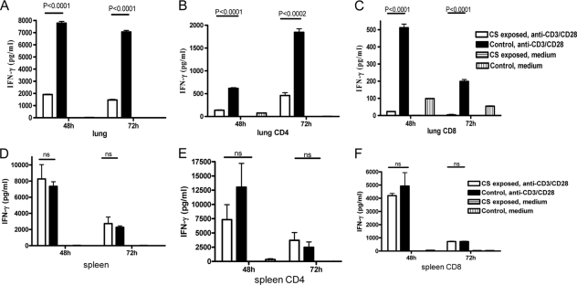
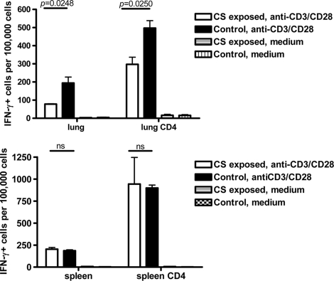
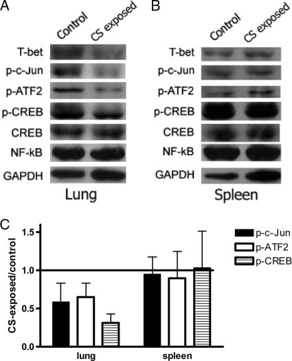
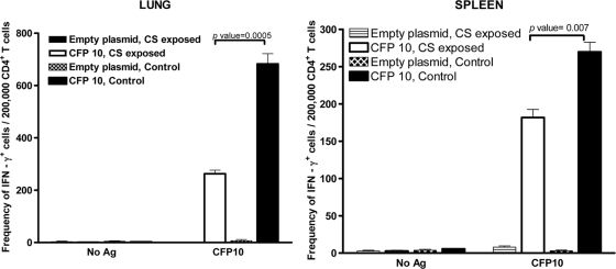
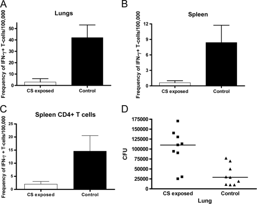
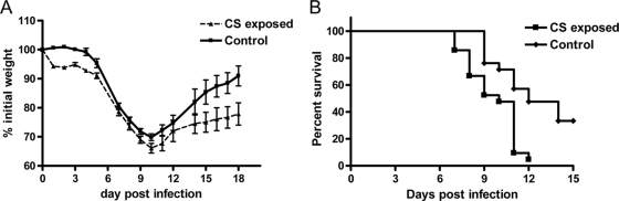
Smoking is associated with increased susceptibility to tuberculosis and influenza. However, little information is available on the mechanisms underlying this increased susceptibility. Mice were left unexposed or were exposed to cigarette smoke and then infected with Mycobacterium tuberculosis by aerosol or influenza A by intranasal infection. Some mice were given a DNA vaccine encoding an immunogenic M. tuberculosis protein. Gamma interferon (IFN-γ) production by T cells from the lungs and spleens was measured. Cigarette smoke exposure inhibited the lung T-cell production of IFN-γ during stimulation in vitro with anti-CD3, after vaccination with a construct expressing an immunogenic mycobacterial protein, and during infection with M. tuberculosis and influenza A virus in vivo. Reduced IFN-γ production was mediated through the decreased phosphorylation of transcription factors that positively regulate IFN-γ expression. Cigarette smoke exposure increased the bacterial burden in mice infected with M. tuberculosis and increased weight loss and mortality in mice infected with influenza virus. This study provides the first demonstration that cigarette smoke exposure directly inhibits the pulmonary T-cell response to M. tuberculosis and influenza virus in a physiologically relevant animal model, increasing susceptibility to both pathogens.
Figures

Similar articles
-
Long-term cigarette smoke exposure dysregulates pulmonary T cell response and IFN-γ protection to influenza virus in mouse.Respir Res. 2021 Apr 20;22(1):112. doi: 10.1186/s12931-021-01713-z. Respir Res. 2021. PMID: 33879121 Free PMC article.
-
CXCR6-Deficiency Improves the Control of Pulmonary Mycobacterium tuberculosis and Influenza Infection Independent of T-Lymphocyte Recruitment to the Lungs.Front Immunol. 2019 Mar 7;10:339. doi: 10.3389/fimmu.2019.00339. eCollection 2019. Front Immunol. 2019. PMID: 30899256 Free PMC article.
-
Expression of interferon-gamma and tumour necrosis factor-alpha messenger RNA does not correlate with protection in guinea pigs challenged with virulent Mycobacterium tuberculosis by the respiratory route.Immunology. 2009 Sep;128(1 Suppl):e296-305. doi: 10.1111/j.1365-2567.2008.02962.x. Epub 2008 Nov 7. Immunology. 2009. PMID: 19016908 Free PMC article.
-
[Novel vaccines against M. tuberculosis].Kekkaku. 2006 Dec;81(12):745-51. Kekkaku. 2006. PMID: 17240920 Review. Japanese.
-
Alteration of the nasal responses to influenza virus by tobacco smoke.Curr Opin Allergy Clin Immunol. 2012 Feb;12(1):24-31. doi: 10.1097/ACI.0b013e32834ecc80. Curr Opin Allergy Clin Immunol. 2012. PMID: 22157158 Free PMC article. Review.
Cited by
-
Heavy Cigarette Smokers in a Chinese Population Display a Compromised Permeability Barrier.Biomed Res Int. 2016;2016:9704598. doi: 10.1155/2016/9704598. Epub 2016 Jun 29. Biomed Res Int. 2016. PMID: 27437403 Free PMC article.
-
Factors associated with smoking among tuberculosis patients in Spain.BMC Infect Dis. 2016 Sep 14;16:486. doi: 10.1186/s12879-016-1819-1. BMC Infect Dis. 2016. PMID: 27629062 Free PMC article.
-
Plasminogen activator inhibitor-1 in cigarette smoke exposure and influenza A virus infection-induced lung injury.PLoS One. 2015 May 1;10(5):e0123187. doi: 10.1371/journal.pone.0123187. eCollection 2015. PLoS One. 2015. PMID: 25932922 Free PMC article.
-
Cigarette smoke increases Staphylococcus aureus biofilm formation via oxidative stress.Infect Immun. 2012 Nov;80(11):3804-11. doi: 10.1128/IAI.00689-12. Epub 2012 Aug 13. Infect Immun. 2012. PMID: 22890993 Free PMC article.
-
Lung Organoids in Smoking Research: Current Advances and Future Promises.Biomolecules. 2022 Oct 12;12(10):1463. doi: 10.3390/biom12101463. Biomolecules. 2022. PMID: 36291672 Free PMC article. Review.
References
-
- Arcavi, L., and N. L. Benowitz. 2004. Cigarette smoking and infection. Arch. Intern. Med. 164:2206-2216. - PubMed
-
- Benowitz, N. L. 2010. Secondhand smoke and infectious disease in adults: a global women's health concern: comment on “passive smoking and tuberculosis.” Arch. Intern. Med. 170:292-293. - PubMed
-
- Boom, W. H. 1996. The role of T-cell subsets in Mycobacterium tuberculosis infection. Infect. Agents Dis. 5:73-81. - PubMed
-
- Brown, D. M., A. M. Dilzer, D. L. Meents, and S. L. Swain. 2006. CD4 T cell-mediated protection from lethal influenza: perforin and antibody-mediated mechanisms give a one-two punch. J. Immunol. 177:2888-2898. - PubMed
-
- Caruso, A. M., N. Serbina, E. Klein, K. Triebold, B. R. Bloom, and J. L. Flynn. 1999. Mice deficient in CD4 T cells have only transiently diminished levels of IFN-gamma, yet succumb to tuberculosis. J. Immunol. 162:5407-5416. - PubMed
Publication types
MeSH terms
Substances
LinkOut - more resources
Full Text Sources
Molecular Biology Databases
