. 2010 Dec 31;285(53):41366-73.
doi: 10.1074/jbc.M110.173815. Epub 2010 Oct 25.

Fission yeast receptor of activated C kinase (RACK1) ortholog Cpc2 regulates mitotic commitment through Wee1 kinase

Affiliations

Fission yeast receptor of activated C kinase (RACK1) ortholog Cpc2 regulates mitotic commitment through Wee1 kinase

Andrés Núñez et al. J Biol Chem. .

Abstract

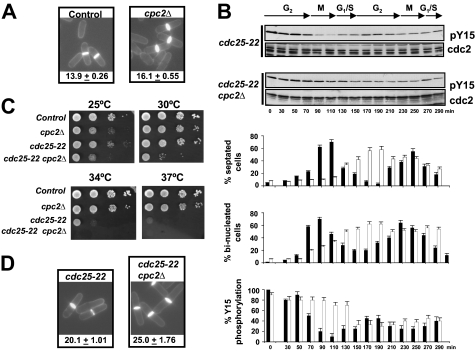
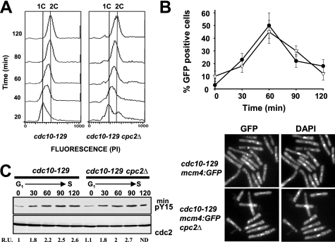
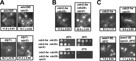
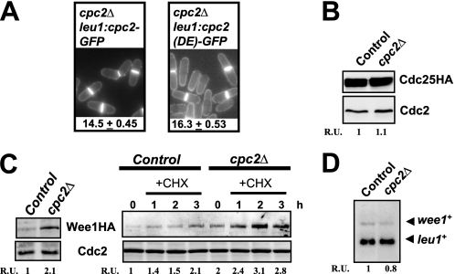
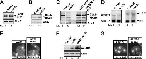
In the fission yeast Schizosaccharomyces pombe, Wee1-dependent inhibitory phosphorylation of the highly conserved Cdc2/Cdk1 kinase determines the mitotic onset when cells have reached a defined size. The receptor of activated C kinase (RACK1) is a scaffolding protein strongly conserved among eukaryotes which binds to other proteins to regulate multiple processes in mammalian cells, including the modulation of cell cycle progression during G(1)/S transition. We have recently described that Cpc2, the fission yeast ortholog to RACK1, controls from the ribosome the activation of MAPK cascades and the cellular defense against oxidative stress by positively regulating the translation of specific genes whose products participate in the above processes. Intriguingly, mutants lacking Cpc2 display an increased cell size at division, suggesting the existence of a specific cell cycle defect at the G(2)/M transition. In this work we show that protein levels of Wee1 mitotic inhibitor are increased in cells devoid of Cpc2, whereas the levels of Cdr2, a Wee1 inhibitor, are down-regulated in the above mutant. On the contrary, the kinetics of G(1)/S transition was virtually identical both in control and Cpc2-less strains. Thus, our results suggest that in fission yeast Cpc2/RACK1 positively regulates from the ribosome the mitotic onset by modulating both the protein levels and the activity of Wee1. This novel mechanism of translational control of cell cycle progression might be conserved in higher eukaryotes.


Figures

FIGURE 1.
FIGURE 1.
Cpc2 is a positive regulator of G2/M transition during the cell cycle. A, cell morphology and size at division (micrometers ± S.D.) in strains MM2 (control) and AN002 (cpc2Δ), growing in YES medium at 28 °C and stained with Calcofluor white. B, cells from strains PPG148 (cdc25-22) and AN-CC18 (cdc25-22 cpc2Δ) were grown to an A600 of 0.3 at 25 °C, shifted to 37 °C for 3.5 h, and then released from the growth arrest by transfer back to 25 °C. Aliquots were taken at different time intervals, and Cdc2 phosphorylation at Tyr15 or total Cdc2 was detected by immunoblotting with anti-Cdc2 pY15 and anti-Cdk1/Cdc2 (PSTAIR) antibodies, respectively. Lower, corresponding percentages of septated cells, binucleated cells, and Tyr15 phosphorylation for cdc25-22 (filled bars) and cdc25–22 cpc2Δ (open bars) cells (n = 4). C, samples containing 104, 103, 102, or 101 cells of strains MM2 (control), PPG148 (cdc25-22), and AN-CC18 (cdc25-22 cpc2Δ) grown in YES medium were spotted onto Bacto Agar supplemented YES plates and incubated for 3 days at either 25 °C, 28 °C, 34 °C, and 37 °C before being photographed. D, cell morphology and size at division in strains PPG148 (cdc25-22) and AN-CC18 (cdc25-22 cpc2Δ) growing at 28 °C determined as described in A.
FIGURE 2.
FIGURE 2.
Cpc2 does not regulate G1/S progression during the cell cycle. A, flow cytometry histograms of cells from strains 326 (cdc10-129) and AN-CC10 (cdc10-129 cpc2Δ) that were synchronized by incubation in YES medium at 37 °C for 3.5 h, transferred back to 25 °C, and incubated for the times indicated. B, upper, cells from strains AN-CC21 (cdc10-129 mcm4:GFP) and AN-CC22 (cdc10-129 mcm4:GFP cpc2Δ) synchronized at G1 by incubation in YES medium at 37 °C for 3.5 h and transferred back to 25 °C. Samples were taken at different times, and the percentage of cells with stable chromatin-bound Mcm4 (GFP-positive cells) was estimated by fluorescence microscopy (n = 3). Filled circles, strain AN-CC21; open triangles, strain AN-CC22. B, lower, micrographs of cells from the above strains expressing Mcm4-GFP taken 60 min after release and stained with DAPI. C, synchronized cells from strains 326 (cdc10-129) and AN-CC10 (cdc10-129 cpc2Δ) released at 25 °C. Aliquots were taken at different time intervals, and Cdc2 phosphorylation at Tyr15 or total Cdc2 was detected by immunoblotting with anti-Cdc2 pY15 or anti-Cdk1/Cdc2 (PSTAIR) antibodies, respectively.
FIGURE 3.
FIGURE 3.
Wee1 is a target for Cpc2 during the regulation of G2/M transition. A, cell morphology and size at division (micrometers ± S.D.) in strains MI709 (Wis1DD) and AN600 (Wis1DD cpc2Δ) (upper), MI204 (sty1Δ) and AN-120 (sty1Δ cpc2Δ) (lower), growing in YES medium after staining with Calcofluor white. B, upper, cell morphology and size at division in strains FY16230 (cdc2-3w cdc25Δ) and AN-CC3 (cdc2-3w cdc25Δ cpc2Δ) prepared as described above. B, lower, samples containing 104, 103, 102, or 101 cells of strains FY16230 (cdc2-3w cdc25Δ) and AN-CC3 (cdc2-3w cdc25Δ cpc2Δ) grown in YES medium were spotted onto Bacto Agar-supplemented YES plates and incubated for 3 days at 25 °C, 28 °C, 34 °C, or 37 °C before being photographed. C, cell morphology and size at division in strains FY7287 (cdc2-1w) and AN-CC1 (cdc2-1w cpc2Δ) (upper), FY7108 (wee1-50) and AN-CC5 (wee1-50 cpc2Δ) (lower) prepared as described above.
FIGURE 4.
FIGURE 4.
Cpc2 negatively regulates Wee1 protein levels. A, ribosome binding of Cpc2 is critical for proper regulation of G2/M transition. Cell morphology and size at division (micrometers ± S.D.) in strains AN071 (cpc2Δ cpc2-GFP) and AN072 (cpc2Δ cpc2 (DE)-GFP) growing in YES medium and stained with Calcofluor white are shown. B, strains FM-23 (cdc25-HA, control) and AN-CC20 (cdc25-HA cpc2Δ) were grown in YES medium to mid log phase. Cdc25 was detected in cell extracts by immunoblotting with anti-HA antibody. Anti-Cdc2 antibody was used for loading control. C, left, strains 1081 (wee1-3HA, control) and AN-CC8 (wee1-3HA cpc2Δ) were grown in YES medium to mid log phase. Total extracts were obtained, and Wee1 was detected by immunoblotting with anti-HA antibody. Anti-Cdc2 antibody was used for loading control. C, right, strains 1081 and AN-CC8 grown in YES medium were treated with 20 μg/ml cycloheximide and aliquots taken at the times indicated. Detection of Wee1 and Cdc2 was performed as described above. D, strains 1081 (control) and AN-CC8 (cpc2Δ) grown in YES medium to early log phase. Total RNA was extracted from each sample and 20 μg resolved in 1.5% agarose-formaldehyde gels. The denatured RNAs were transferred to nylon membranes and hybridized with 32P-labeled probes for wee1+ and leu1+ (loading control).
FIGURE 5.
FIGURE 5.
Protein level of the Wee1 inhibitor Cdr2 is positively regulated by Cpc2. A, strains MM-76 (pom1-GFP, control) and AN-CC35 (pom1-GFP cpc2Δ) were grown in YES medium to mid log phase, total cell extracts were obtained, and Pom1 was detected by immunoblotting with anti-GFP antibody. Anti-Cdc2 antibody was used for loading control. B, strains LW117 (nim1-HA6H, control) and AN-CC15 (nim1-HA6H cpc2Δ) were grown as above described, and Nim1 was detected by immunoblotting with anti-HA antibody. C, strains JK2310 (cdr2-HA6H, control), AN-CC14 (cdr2-HA6H cpc2Δ), AN-CC23 (cdr2-HA6H cpc2-GFP), and AN-CC24 (cdr2-HA6H cpc2(DE)-GFP) were grown in YES medium as above, and Cdr2 was detected by immunoblotting with anti-HA antibody. D, strains LW117 (control), AN-CC15 (cpc2Δ), JK2310 (control), and AN-CC14 (cdr2-HA6H cpc2Δ) were grown in YES medium to early log phase. Total RNA was extracted from each sample and 20 μg resolved in 1.5% agarose-formaldehyde gels. The denatured RNAs were transferred to nylon membranes and hybridized with 32P-labeled probes for nim1+ (strains LW117 and AN-CC15), cdr2+ (strains JK2310 and AN-CC14), and leu1+ (loading control). E, cell morphology and size at division (micrometers ± S.D.) in strains JK2240 (cdr2Δ) and AN-CC12 (cdr2Δ cpc2Δ) growing in YES medium and stained with Calcofluor white are shown. F, strains 1081 (wee1-3HA, control), AN-CC45 (wee1-3HA cdr2Δ), and AN-CC46 (wee1-3HA cdr2Δ cpc2Δ) were grown in YES medium to mid log phase. Total extracts were obtained, and Wee1 was detected by immunoblotting with anti-HA antibody. Anti-Cdc2 antibody was used for loading control. G, cell morphology and size at division (micrometers ± S.D.) in strains TS313 (pom1Δ) and AN-CC27 (pom1Δ cpc2Δ) were determined as described in A.

Similar articles

Cited by

References

    1. Nurse P. (1990) Nature 344, 503–508 - PubMed
    1. Rupes I. (2002) Trends Genet. 18, 479–485 - PubMed
    1. Russell P., Nurse P. (1987) Cell 49, 559–567 - PubMed
    1. Gould K. L., Nurse P. (1989) Nature 342, 39–45 - PubMed
    1. Featherstone C., Russell P. (1991) Nature 349, 808–811 - PubMed

Publication types

MeSH terms

LinkOut - more resources