. 2010 Dec 31;285(53):41491-500.
doi: 10.1074/jbc.M110.153338. Epub 2010 Oct 25.

Eosinophil cysteinyl leukotriene synthesis mediated by exogenous secreted phospholipase A2 group X

Affiliations

Eosinophil cysteinyl leukotriene synthesis mediated by exogenous secreted phospholipase A2 group X

Ying Lai et al. J Biol Chem. .

Abstract

Secreted phospholipase A(2) group X (sPLA(2)-X) has recently been identified in the airways of patients with asthma and may participate in cysteinyl leukotriene (CysLT; C(4), D(4), and E(4)) synthesis. We examined CysLT synthesis and arachidonic acid (AA) and lysophospholipid release by eosinophils mediated by recombinant human sPLA(2)-X. We found that recombinant sPLA(2)-X caused marked AA release and a rapid onset of CysLT synthesis in human eosinophils that was blocked by a selective sPLA(2)-X inhibitor. Exogenous sPLA(2)-X released lysophospholipid species that arise from phospholipids enriched in AA in eosinophils, including phosphatidylcholine, phosphatidylinositol, and phosphatidylethanolamine as well as plasmenyl phosphatidylcholine and phosphatidylethanolamine. CysLT synthesis mediated by sPLA(2)-X but not AA release could be suppressed by inhibition of cPLA(2)α. Exogenous sPLA(2)-X initiated Ser(505) phosphorylation of cPLA(2)α, an intracellular Ca(2+) flux, and translocation of cPLA(2)α and 5-lipoxygenase in eosinophils. Synthesis of CysLTs in response to sPLA(2)-X or lysophosphatidylcholine was inhibited by p38 or JNK inhibitors but not by a MEK 1/2 inhibitor. A further increase in CysLT synthesis was induced by the addition of sPLA(2)-X to eosinophils under conditions of N-formyl-methionyl-leucyl-phenylalanine-mediated cPLA(2)α activation. These results indicate that sPLA(2)-X participates in AA and lysophospholipid release, resulting in CysLT synthesis in eosinophils through a mechanism involving p38 and JNK MAPK, cPLA(2)α, and 5-lipoxygenase activation and resulting in the amplification of CysLT synthesis during cPLA(2)α activation. Transactivation of eosinophils by sPLA(2)-X may be an important mechanism leading to CysLT formation in the airways of patients with asthma.


Figures

FIGURE 1.
FIGURE 1.
Time course and concentration dependence of sPLA2-X mediated eicosanoid synthesis by eosinophils. A, following treatment with 100 nm exogenous sPLA2-X, there was an increase in [3H]AA release by eosinophils over time that reached a plateau 15 min after addition of the enzyme (p < 0.0001). The [3H]AA release is expressed in terms of percentage of total [3H]AA incorporated. B, eosinophil synthesis of CysLTs increased over time after treatment with 100 nm exogenous sPLA2-X, reaching a maximum 20 min after the addition of the enzyme (p = 0.002). C, the levels of CysLTs following treatment of eosinophils for 20 min with buffer control or 1, 10, or 100 nm sPLA2-X increased with increasing concentrations of the recombinant enzyme (p = 0.008). Error bars, S.E.
FIGURE 2.
FIGURE 2.
Effects of sPLA2 inhibitors on AA release and CysLT synthesis from sPLA2-X-treated eosinophils. A, eosinophils treated with 100 nm exogenous sPLA2-X (black bar) had significant AA release relative to buffer control (white bar; *, p = 0.01). The sPLA2-X-specific inhibitor (ROC-0929) caused a dose-dependent decrease in [3H]AA release by eosinophils following treatment with 100 nm exogenous sPLA2-X at inhibitor concentrations ranging from 10 to 1000 nm (p < 0.0001). B, eosinophils also had a significant increase in CysLT synthesis after treatment with 100 nm exogenous sPLA2-X (black bar) relative to buffer control (white bar; †, p = 0.002). The synthesis of CysLTs by eosinophils after treatment with 100 nm sPLA2-X was inhibited in a dose-dependent manner by increasing concentrations of the ROC-0929 inhibitor ranging from 1 to 100 nm (p = 0.002). Treatment with a structurally related inhibitor devoid of sPLA2-X inhibitory activity (ROC-0428) had no effect on sPLA2-X-mediated CysLT synthesis. Eosinophils did not synthesize CysLTs in response to 100 nm sPLA2-X that had been heated for 10 min (Denatured). Error bars, S.E.
FIGURE 3.
FIGURE 3.
Exogenous sPLA2-X-mediated generation of lysophospholipids by eosinophils. A–D, treatment of eosinophils with sPLA2-X (100 nm) initiated the generation of LysoPC species (A), lysophosphatidylethanolamine (LysoPE) species (B), lysophosphatidylserine (LysoPS) species (C), and lysophosphatidylinositol (LysoPI) species (D) relative to control conditions (white bar) and relative to eosinophils treated with fMLP (100 nm). *, p ≤ 0.01 and †, p ≤ 0.05 overall. ‡, p ≤ 0.01 and §, p ≤ 0.05 versus fMLP. The complete analysis of lysophospholipid species is presented in supplemental Figs. 3 and 4. Error bars, S.E.
FIGURE 4.
FIGURE 4.
Effects of sPLA2-X mediated by cPLA2α in eosinophils. A, treatment of eosinophils with 100 nm exogenous sPLA2-X for 20 min (black bar) increased [3H]AA release over buffer control (white bar; *, p = 0.001). The cPLA2α inhibitors Pyr-2 (5 and 10 μm) and Wyeth-2 (5 μm) failed to significantly decrease sPLA2-X-mediated [3H]AA release by eosinophils (p = 0.44). B, in contrast to AA release, the significant increase in CysLT synthesis by eosinophils following treatment with 100 nm sPLA2-X (†, p = 0.02) was inhibited in a dose-dependent manner by Pyr-2 in concentrations ranging from 1 to 10 μm (p = 0.007) and by 5 μm Wyeth-2. C, Western blots of cell lysates from eosinophils treated with buffer control (Ctrl), 100 nm fMLP, or 100 nm sPLA2-X demonstrate phosphorylation of cPLA2α at Ser505 relative to total cPLA2α. An example blot from one of the three replicate blots from different subjects is shown at the bottom. D, cell-wide changes in cytoplasmic Ca2+ concentration ([Ca2+]i) were monitored by fluo4-AM-loaded eosinophils, reported as the fluorescence intensity at each time point relative to the resting fluorescence (F/F0). Relative to the buffer control (a), the addition of sPLA2-X (b; 100 nm) caused an increase in [Ca2+]i that was further increased by a higher concentration of sPLA2-X (c; 200 nm) and fMLP (d; 100 nm). Ionophore (e; A23187, 10 μm) caused a sustained increase in [Ca2+]i. Images of the intracellular fluorescence at each point are shown at the top of the plot. Error bars, S.E.
FIGURE 5.
FIGURE 5.
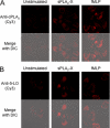
Translocation of cPLA2 and 5-LO in response to sPLA2-X. Eosinophils were allowed to adhere to BSA-coated coverslips and treated with vehicle alone (Unstimulated), sPLA2-X (100 nm), or fMLP (100 nm). The fMLP-stimulated cells served as a positive control. Cells were fixed, permeabilized, and immunostained with antibodies directed against cPLA2 (A) and 5-LO (B). Fluorescence from the secondary Cy3-labeled antibody was visualized with confocal microscopy. A, in unstimulated cells, the cPLA2 immunostaining was faint and diffuse, but the immunostaining increased following treatment of the cells with sPLA2-X and localized in the cells in the perinuclear space as well as punctate cytoplasmic staining. Similar immunostaining was observed for eosinophils treated with fMLP as a positive control. B, the 5-LO immunostaining in unstimulated cells was also faint and diffuse, but the immunostaining increased following treatment of the cells with sPLA2-X in both perinuclear and focal intracytoplasmic locations. Similar immunostaining for 5-LO was observed for eosinophils treated with fMLP as a positive control.
FIGURE 6.
FIGURE 6.
Effects of kinase inhibitors on eosinophil CysLT synthesis. A, relative to the maximum amount of CysLT generated by treatment of eosinophils with sPLA2-X (100 nm) (black bar), CysLT synthesis was not inhibited by a MEK 1/2 inhibitor (U0126, 10 μm) but was significantly inhibited by a p38 inhibitor (SB203580, 30 μm) and by a JNK inhibitor (SP600125, 20 μm). B, relative to maximum CysLT synthesis by eosinophils treated with LysoPC (10 μm) (black bar), CysLT synthesis was not inhibited by a MEK 1/2 inhibitor (U0126, 10 μm) but was significantly inhibited by p38 (SB203580, 30 μm) and JNK inhibitors (SP600125, 20 μm). C, eosinophil CysLT synthesis after treatment with fMLP (100 nm) (black bar) was individually inhibited by MEK 1/2 (U0126, 10 μm), p38 (SB203580, 30 μm), and JNK (SP600125, 20 μm) inhibitors. *, p = not significant; †, p < 0.05; §, p < 0.01. Error bars, S.E.
FIGURE 7.
FIGURE 7.
Activation of CysLT synthesis in fMLP-stimulated eosinophils by sPLA2-X. A, in comparison with unstimulated eosinophils (Ctrl), eosinophils treated with fMLP (10 nm) had an increase in CysLT synthesis that was further increased by the addition of sPLA2-X at concentrations of 10 and 100 nm (p = 0.01). B, at a higher concentration of fMLP (100 nm), eosinophil CysLT synthesis was also further augmented by sPLA2-X at concentrations of 10 and 100 nm (p = 0.03). Additional plots of the individual data from each eosinophil donor are shown in supplemental Fig. 2. Error bars, S.E.
FIGURE 8.
FIGURE 8.
Schematic representation of the events during sPLA2-X-mediated CysLT synthesis by eosinophils. sPLA2-X causes the release of lysophospholipids (LysoPL) and free fatty acids (FFA), including AA from phospholipid species enriched in AA. sPLA2-X causes CysLT synthesis that is dependent upon cPLA2α and initiates a Ca2+ flux and cPLA2α phosphorylation in eosinophils. Prior research has shown that LysoPC causes a Ca2+ flux in human eosinophils. We found that the sPLA2-X causes a Ca2+ flux and that sPLA2-X- and LysoPC-induced CysLT synthesis could be inhibited by p38 and JNK inhibitors but not by a MEK 1/2 inhibitor. Free AA released by sPLA2-X may contribute to additional CysLT synthesis based on the observation that the addition of sPLA2-X to eosinophils treated with fMLP leads to additional CysLT synthesis.

Similar articles

Cited by

References

    1. Hallstrand T. S., Henderson W. R., Jr. (2010) Curr. Opin. Allergy Clin. Immunol. 10, 60–66 - PMC - PubMed
    1. Hallstrand T. S., Moody M. W., Aitken M. L., Henderson W. R., Jr. (2005) J. Allergy Clin. Immunol. 116, 586–593 - PMC - PubMed
    1. Pavord I. D., Ward R., Woltmann G., Wardlaw A. J., Sheller J. R., Dworski R. (1999) Am. J. Respir Crit. Care Med. 160, 1905–1909 - PubMed
    1. Romagnoli M., Vachier I., Tarodo de la Fuente P., Meziane H., Chavis C., Bousquet J., Godard P., Chanez P. (2002) Eur. Respir. J. 20, 1370–1377 - PubMed
    1. Ago H., Kanaoka Y., Irikura D., Lam B. K., Shimamura T., Austen K. F., Miyano M. (2007) Nature 448, 609–612 - PubMed

Publication types

MeSH terms