

Vitamin D suppresses Th17 cytokine production by inducing C/EBP homologous protein (CHOP) expression
- PMID: 20974859
- PMCID: PMC2998156
- DOI: 10.1074/jbc.C110.185777
Vitamin D suppresses Th17 cytokine production by inducing C/EBP homologous protein (CHOP) expression
Abstract
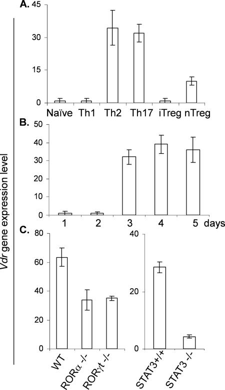
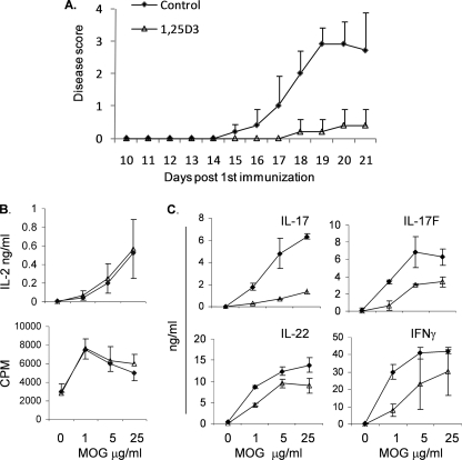
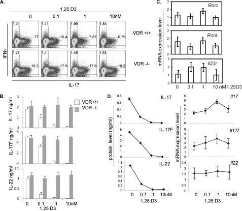
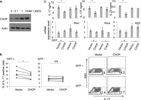
Vitamin D has been shown to have immunomodulatory function, but the molecular basis for it has not been well understood. In this study, we found that vitamin D receptor expression was induced in a CD4+ effector T cell lineage, Th17 cells, which required the transcription factors, RORα, RORγt, and STAT3. Treatment of mice with an active ligand of vitamin D receptor (VDR), 1,25-dihydroxyvitamin D(3) (1,25D3), ameliorated experimental autoimmune encephalomyelitis, accompanied with reduced IL-17 and IL-17F expression. In vitro, treatment of CD4+ T cells with the physiological doses of 1,25D3 preferentially inhibited cytokine production by Th17 cells, in a VDR-dependent manner, without affecting the expression of transcription factors or surface molecules. Moreover, at these concentrations, cytokine expression was suppressed only at protein but not at mRNA levels. Stimulation of Th17 cells with 1,25D3, in a concentration-dependent manner, induced the expression of C/EBP homologous protein (CHOP), a molecule involved in endoplasmic reticulum stress and translational inhibition. In addition, overexpression of CHOP in developing Th17 cells suppressed their cytokine production. Our results suggest a novel, post-transcriptional mechanism whereby Th17 cytokines are inhibited by VDR, which may underscore future therapeutic usage of vitamin D in treatment of autoimmune diseases.
Figures
Similar articles
-
1α,25-Dihydroxyvitamin D3 and all-trans retinoic acid synergistically inhibit the differentiation and expansion of Th17 cells.Immunol Lett. 2010 Nov 30;134(1):7-16. doi: 10.1016/j.imlet.2010.07.002. Epub 2010 Jul 23. Immunol Lett. 2010. PMID: 20655952
-
1,25-dihydroxyvitamin D(3) ameliorates Th17 autoimmunity via transcriptional modulation of interleukin-17A.Mol Cell Biol. 2011 Sep;31(17):3653-69. doi: 10.1128/MCB.05020-11. Epub 2011 Jul 11. Mol Cell Biol. 2011. PMID: 21746882 Free PMC article.
-
Converging pathways lead to overproduction of IL-17 in the absence of vitamin D signaling.Int Immunol. 2011 Aug;23(8):519-28. doi: 10.1093/intimm/dxr045. Epub 2011 Jun 22. Int Immunol. 2011. PMID: 21697289 Free PMC article.
-
New understanding of the molecular mechanism of receptor-mediated genomic actions of the vitamin D hormone.Bone. 1995 Aug;17(2 Suppl):33S-38S. doi: 10.1016/8756-3282(95)00205-r. Bone. 1995. PMID: 8579895 Review.
-
The vitamin D hormone and its nuclear receptor: molecular actions and disease states.J Endocrinol. 1997 Sep;154 Suppl:S57-73. J Endocrinol. 1997. PMID: 9379138 Review.
Cited by
-
Effects of 1,25-dihydroxyvitamin D3 on IL-17/IL-23 axis, IFN-γ and IL-4 expression in systemic lupus erythematosus induced mice model.Iran J Basic Med Sci. 2016 Apr;19(4):374-80. Iran J Basic Med Sci. 2016. PMID: 27279980 Free PMC article.
-
Psoriasis: Are Your Patients D-pleted? A Brief Literature Review on Vitamin D Deficiency and Its Role in Psoriasis.J Clin Aesthet Dermatol. 2022 Mar;15(3 Suppl 1):S30-S33. J Clin Aesthet Dermatol. 2022. PMID: 35382439 Free PMC article. Review.
-
Differential effect of vitamin D on NOD2- and TLR-induced cytokines in Crohn's disease.Mucosal Immunol. 2014 Nov;7(6):1405-15. doi: 10.1038/mi.2014.30. Epub 2014 Apr 30. Mucosal Immunol. 2014. PMID: 24781050
-
The role of human milk nutrients in preventing necrotizing enterocolitis.Front Pediatr. 2023 Jun 2;11:1188050. doi: 10.3389/fped.2023.1188050. eCollection 2023. Front Pediatr. 2023. PMID: 37334221 Free PMC article. Review.
-
The Impact of Vitamin D in the Prevention of Influenza, COVID-19, and Dengue: A Review.Biomedicines. 2025 Apr 9;13(4):927. doi: 10.3390/biomedicines13040927. Biomedicines. 2025. PMID: 40299497 Free PMC article. Review.
References
Publication types
MeSH terms
Substances
Grants and funding
LinkOut - more resources
Full Text Sources
Other Literature Sources
Medical
Research Materials
Miscellaneous
