

Immunocytochemistry for amoxicillin and its use for studying uptake of the drug in the intestine, liver, and kidney of rats
- PMID: 20974868
- PMCID: PMC3019652
- DOI: 10.1128/AAC.01031-10
Immunocytochemistry for amoxicillin and its use for studying uptake of the drug in the intestine, liver, and kidney of rats
Abstract
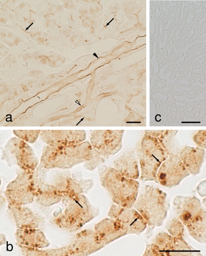
Specific transport systems for penicillins have been recognized, but their in vivo role in the context of other transporters remains unclear. We produced a serum against amoxicillin (anti-AMPC) conjugated to albumin with glutaraldehyde. The antiserum was specific for AMPC and ampicillin (ABPC) but cross-reacted weakly with cephalexin. This enabled us to develop an immunocytochemical (ICC) method for detecting the uptake of AMPC in the rat intestine, liver, and kidney. Three hours after a single oral administration of AMPC, the ICC method revealed that AMPC distributed to a high degree in the microvilli, nuclei, and cytoplasm of the absorptive epithelial cells of the intestine. AMPC distributed in the cytoplasm and nuclei of the hepatocytes in a characteristic granular morphology on the bile capillaries, and in addition, AMPC adsorption was observed on the luminal surface of the capillaries, intercalated portions, and interlobular bile ducts on the bile flow. Almost no AMPC could be detected 6 h postadministration in either the intestine or the liver. Meanwhile, in the kidney, AMPC persisted until 12 h postadministration to a high degree in the proximal tubules, especially in the S3 segment cells in the tubular lumen, in which numerous small bodies that strongly reacted with the antibody were observed. All these sites of AMPC accumulation correspond well to specific sites where certain transporter systems for penicillins occur, suggesting that AMPC is actually and actively absorbed, eliminated, or excreted at these sites, possibly through such certain penicillin transporters.
Figures


Similar articles
-
Immunocytochemistry for bestatin and its application to drug accumulation studies in rat intestine and kidney.J Mol Histol. 2011 Dec;42(6):589-96. doi: 10.1007/s10735-011-9365-z. Epub 2011 Oct 18. J Mol Histol. 2011. PMID: 22006287
-
An in vivo role of Mrp2 in the rat hepatocytes by immunocytochemistry for amoxicillin using the transporter-deficient EHBR.J Mol Histol. 2012 Jun;43(3):371-8. doi: 10.1007/s10735-012-9406-2. Epub 2012 Mar 25. J Mol Histol. 2012. PMID: 22446938
-
Immunocytochemistry for vancomycin using a monoclonal antibody that reveals accumulation of the drug in rat kidney and liver.Antimicrob Agents Chemother. 2012 Nov;56(11):5883-91. doi: 10.1128/AAC.01267-12. Epub 2012 Sep 4. Antimicrob Agents Chemother. 2012. PMID: 22948874 Free PMC article.
-
[Distribution and accumulation of antibiotics in cells and tissues and toxicity studies by immunocytochemistry].Yakugaku Zasshi. 2011;131(6):949-60. doi: 10.1248/yakushi.131.949. Yakugaku Zasshi. 2011. PMID: 21628984 Review. Japanese.
-
Drug transport in intestine, liver and kidney.Arch Toxicol. 1987;60(1-3):37-42. doi: 10.1007/BF00296944. Arch Toxicol. 1987. PMID: 3304216 Review.
Cited by
-
Immunocytochemistry for bestatin and its application to drug accumulation studies in rat intestine and kidney.J Mol Histol. 2011 Dec;42(6):589-96. doi: 10.1007/s10735-011-9365-z. Epub 2011 Oct 18. J Mol Histol. 2011. PMID: 22006287
-
Study of protein haptenation by amoxicillin through the use of a biotinylated antibiotic.PLoS One. 2014 Mar 3;9(3):e90891. doi: 10.1371/journal.pone.0090891. eCollection 2014. PLoS One. 2014. PMID: 24595455 Free PMC article.
-
Immunohistochemistry for Anti-diabetes Drug, Alogliptin Using a Newly Prepared Monoclonal Antibody: Its Precise Localization in Rat Small Intestine.Acta Histochem Cytochem. 2019 Feb 28;52(1):27-34. doi: 10.1267/ahc.18036. Epub 2019 Feb 23. Acta Histochem Cytochem. 2019. PMID: 30923413 Free PMC article.
-
Immunohistochemical Localization of Alogliptin, a DPP-4 Inhibitor, in Tissues of Normal and Type 2 Diabetes Model Rat.Acta Histochem Cytochem. 2022 Dec 28;55(6):185-192. doi: 10.1267/ahc.22-00032. Epub 2022 Dec 15. Acta Histochem Cytochem. 2022. PMID: 36688140 Free PMC article.
-
An in vivo role of Mrp2 in the rat hepatocytes by immunocytochemistry for amoxicillin using the transporter-deficient EHBR.J Mol Histol. 2012 Jun;43(3):371-8. doi: 10.1007/s10735-012-9406-2. Epub 2012 Mar 25. J Mol Histol. 2012. PMID: 22446938
References
-
- Adibi, S. A. 2003. Regulation of expression of the intestinal oligopeptide transporter (Pept-1) in health and disease. Am. J. Physiol. Gastrointest. Liver Physiol. 285:G779-G788. - PubMed
-
- Apiwattanakul, N., T. Sekine, A. Chairoungdua, Y. Kanai, N. Nakajima, S. Sophasan, and H. Endou. 1999. Transport properties of nonsteroidal anti-inflammatory drugs by organic anion transporter 1 expressed in Xenopus laevis oocytes. Mol. Pharmacol. 55:847-854. - PubMed
-
- Barza, N., J. Brush, N. C. Bergeron, O. Kemmotsu, and L. Weinstein. 1975. Extraction of antibiotics from the circulation by liver and kidney: effect of probenecid. J. Infect. Dis. 131:S86-S97. - PubMed
-
- Bergwerk, A. J., K. Shi, A. C. Ford, N. Kanai, E. Jacquemin, R. D. Burk, S. Bai, P. R. Novikoff, B. Stieger, P. J. Meier, V. L. Schuster, and A. W. Wolkoff. 1996. Immunologic distribution of an organic anion transport protein in rat liver and kidney. Am. J. Physiol. 271:G231-G238. - PubMed
Publication types
MeSH terms
Substances
LinkOut - more resources
Full Text Sources
