

doi: 10.1104/pp.110.164814.
Epub 2010 Oct 25.
Advancing our understanding and capacity to engineer nature's CO2-sequestering enzyme, Rubisco
Affiliations
- PMID: 20974895
- PMCID: PMC3075749
- DOI: 10.1104/pp.110.164814
Item in Clipboard
Advancing our understanding and capacity to engineer nature's CO2-sequestering enzyme, Rubisco
Plant Physiol.
2011 Jan.
No abstract available
Figures
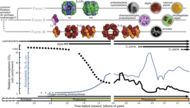
Hypothetical profiles of Rubisco phylogeny, the evolutionary timelines of different photosynthetic organisms, and variation in atmospheric CO2 (thicker line) and O2 levels during earth’s history. Hypothetical atmospheric CO2 and O2 levels prior to 0.6 billion years ago are represented by dotted lines. Quaternary structures of each Rubisco were drawn with Pymol using Protein Data Bank coordinates for the spinach (Spinacia oleracea) (L2)4S8 (8RUC), R. rubrum L2 (5RUB), Pyrococcus horikoshii (L2)4 (2CWX), and Thermococcus kodakaraensis (L2)5 (1GEH) enzymes. Structures for larger form II (L2)n Rubiscos are unavailable. Circular images depict types of organisms where the different Rubisco forms are found. Figure details were adapted from Tabita et al. (2008) and Badger et al. (2002).
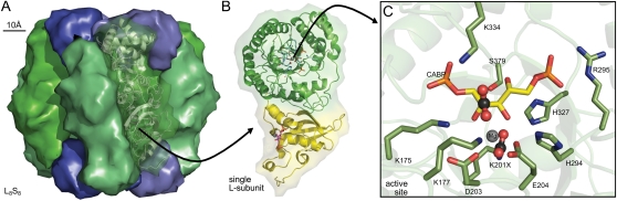
Conserved structural features of Rubisco. A, Spinach L8S8 Rubisco (Protein Data Bank 8RUC) drawn using Pymol to highlight arrangement of the S-subunits (blue) capping the catalytic core of four L2 subunits (green). B, Structural details for one L-subunit of an L2 pair highlighting one active site within the α/β-barrel of the C-terminal domain (green ribbons) and residues in the N-terminal domain (yellow ribbons) that contribute to the second active site in each L2. C, Arrangement of the conserved Rubisco active-site residues within a L-subunit C-terminal domain relative to carbamylated Lys-201 (K201X), bound Mg2+, and the six-carbon reaction intermediate mimic, 2-carboxyarabinitol 1,5-bisphosphate (CABP). The activating CO2 in K201X and the approximate positioning of substrate CO2 that binds to C-2 of the RuBP enediol are highlighted in ball-and-stick representations. Residues are numbered relative to spinach Rubisco. The conserved active site residues Glu-60 and Asn-123 from the N-terminal domain of the paired L-subunit are not shown.
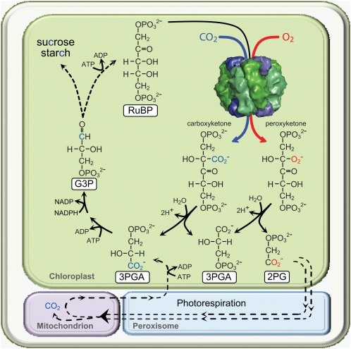
Simplified scheme illustrating how CO2 fixed to RuBP by Rubisco is distributed among the resulting two molecules of 3PGA that feed into the photosynthetic Calvin cycle to produce triose phosphates (glyceraldehyde 3-phosphate [G3P]) for carbohydrate synthesis or RuBP regeneration. The contrasting oxygenation reaction of Rubiscos produces 2-phosphoglycolate (2PG), which requires the photorespiratory pathway to recycle it back to 3PGA. Photorespiration is a complex pathway that involves four subcellular compartments and multiple enzymatic steps (represented by dashed lines), requires additional energy (ATP), and results in a loss of fixed CO2 in the mitochondria (Maurino and Peterhansel, 2010).
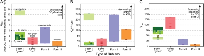
Comparative catalytic features of different Rubisco forms measured at 25°C. Individual dashes in each column represent separate catalytic measurements for each Rubisco form (detailed in Supplemental Table S1 ). Yellow circles indicate the catalytic measurements for green algal Rubisco. SC/O values are calculated as (vCO2/KmO2)/(vO2/KmCO2), where vCO2 and vO2 are the maximum rates of RuBP carboxylation and oxygenation and KmO2 and KmCO2 are the apparent Km values for O2 and CO2, respectively.
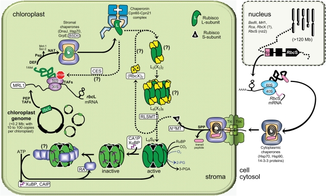
The complexity of Rubisco biogenesis and its regulation by RA in vascular plant chloroplasts. Putative and known Rubisco-specific processes and interacting molecular partners (coded by genes in the nucleus as shown) are highlighted in white rectangles. See text for details of the processes, abbreviations, and the challenges faced in modifying the L8S8 enzyme. Figure details were adapted from Nishimura et al. (2008). Uncertainties in the biogenesis process are indicated by question marks. Hsp, Heat shock proteins; PEP, plastid-encoded RNA polymerase proteins; Pep, unknown Met-1, Ser-2 peptidase activity; Rca, nucleus gene coding RA; TAFs, nucleus-encoded trans-acting factors, Tic and Toc, chloroplast translocon inner and outer membrane complexes respectively; 30S/50S and 40S/60S, stromal and cytosolic ribosomal subunits.
References
-
- Andersson I, Backlund A. (2008) Structure and function of Rubisco. Plant Physiol Biochem 46: 275–291 - PubMed
-
- Badger MR, Hanson D, Price GD. (2002) Evolution and diversity of CO2 concentrating mechanisms in cyanobacteria. Funct Plant Biol 29: 161–173 - PubMed
-
- Bershtein S, Tawfik DS. (2008) Advances in laboratory evolution of enzymes. Curr Opin Chem Biol 12: 151–158 - PubMed
Publication types
MeSH terms
Substances
LinkOut - more resources
Full Text Sources
Other Literature Sources
