. 2011 Jan;21(1):12-20.
doi: 10.1101/gr.108696.110. Epub 2010 Oct 25.

OPA1 links human mitochondrial genome maintenance to mtDNA replication and distribution

Affiliations

OPA1 links human mitochondrial genome maintenance to mtDNA replication and distribution

Ghizlane Elachouri et al. Genome Res. 2011 Jan.

Abstract

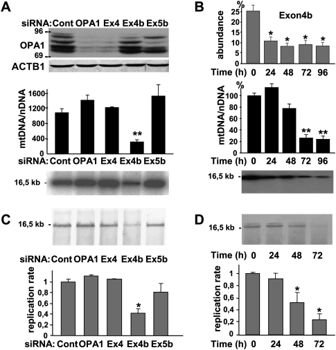
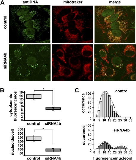
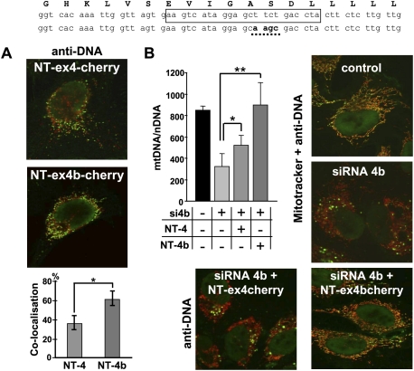
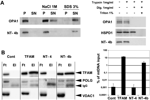
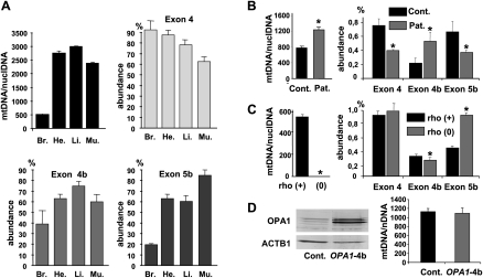
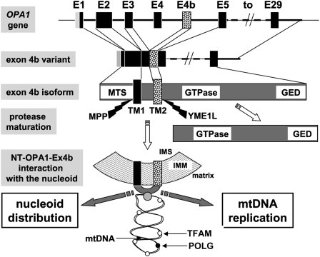
Eukaryotic cells harbor a small multiploid mitochondrial genome, organized in nucleoids spread within the mitochondrial network. Maintenance and distribution of mitochondrial DNA (mtDNA) are essential for energy metabolism, mitochondrial lineage in primordial germ cells, and to prevent mtDNA instability, which leads to many debilitating human diseases. Mounting evidence suggests that the actors of the mitochondrial network dynamics, among which is the intramitochondrial dynamin OPA1, might be involved in these processes. Here, using siRNAs specific to OPA1 alternate spliced exons, we evidenced that silencing of the OPA1 variants including exon 4b leads to mtDNA depletion, secondary to inhibition of mtDNA replication, and to marked alteration of mtDNA distribution in nucleoid and nucleoid distribution throughout the mitochondrial network. We demonstrate that a small hydrophobic 10-kDa peptide generated by cleavage of the OPA1-exon4b isoform is responsible for this process and show that this peptide is embedded in the inner membrane and colocalizes and coimmunoprecipitates with nucleoid components. We propose a novel synthetic model in which a peptide, including two trans-membrane domains derived from the N terminus of the OPA1-exon4b isoform in vertebrates or from its ortholog in lower eukaryotes, might contribute to nucleoid attachment to the inner mitochondrial membrane and promotes mtDNA replication and distribution. Thus, this study places OPA1 as a direct actor in the maintenance of mitochondrial genome integrity.


Figures

Figure 1.
Figure 1.
Mitochondrial genome maintenance and replication require OPA1-exon4b. (A) Western blot using OPA1 and ACTB1 antibodies of protein extracts (top), and evaluation by Q-PCR (middle) and Southern blot (bottom) of mtDNA copy number from control HeLa cells (Cont) or cells transfected for 72 h with siRNAs targeting all OPA1 mRNA (OPA1), or those including exon 4 (Ex4), 4b (Ex4b), or 5b (Ex5b), show a specific mtDNA loss in cells silenced for OPA1-exon4b transcripts. Note that because OPA1 isoforms including the exon 4b domain account for <25% of all OPA1 isoforms, their silencing can barely be detected by Western blot. (n = 6, one-way ANOVA; [**] P-value inferior to 0.001). (B) Quantification of the relative abundances of OPA1 transcripts including exon 4b (top) and of mtDNA in HeLa cells transfected by the exon 4b siRNA during a 24-h period time course reveals a delay between OPA1-exon4b silencing and the mtDNA loss, without evidencing mtDNA degradation or deletions on the Southern blot (bottom). (n = 3, one-way ANOVA; P-value inferior to 0.01 [*] and 0.001 [**]). (C) Southern–Western blot using anti-BrdU antibodies on DNA extracts (top) and quantification of the replication rate (bottom) in control HeLa cells (Cont) or cells transfected for 48 h with the siRNAs targeting all OPA1 mRNA (OPA1) or those including exon 4 (Ex4), 4b (Ex4b), or 5b (Ex5b) shows a significant 60% decrease of BrdU incorporation in mtDNA, in cells silenced for OPA1-exon4b (n = 3, one-way ANOVA; [*] P-value inferior to 0.01). (D) Southern–Western blot of DNA extracts (top) and quantification of the replication rate (bottom) from HeLa cells transfected with the exon 4b siRNA for the indicated times illustrates a progressive inhibition of mtDNA replication starting readily after OPA1-exon4b silencing (n = 3, one-way ANOVA; [*] P-value inferior to 0.01).
Figure 2.
Figure 2.
mtDNA and nucleoid distributions are dismantled by OPA1-exon4b silencing. (A) Fluorescence images using anti-DNA and Mitotracker of HeLa cells (control, top) and cells transfected with the exon 4b siRNA (siRNA4b, bottom) reveal a dramatic change in mtDNA distribution in cells silenced for exon 4b, whereas the mitochondrial network remains tubular as in control cells. (B) Quantifications of cytoplasmic anti-DNA fluorescence (top, relative units) and of the number of nucleoids per cell (bottom) in control (Cont) and exon 4b siRNA-transfected cells (si4b) disclose a decrease in mtDNA-associated fluorescence (50%) and in the number of nucleoids (60%), in exon 4b–silenced cells (n = 8; Student's t-test; [*] P-value inferior to 0.01). (C) Distribution of the anti-DNA fluorescence per nucleoid (relative units) in control (Control, top) and exon 4b siRNA-transfected (si4b, bottom) cells (n = 8) reveals a population with higher than normal fluorescence levels in exon 4b–silenced cells.
Figure 3.
Figure 3.
OPA1-exon4b N terminus colocalizes with mtDNA and rescues exon 4b silencing. The human exon 4b peptide sequence (top) stands above the corresponding wild-type and mutagenized exon 4b nucleotide sequences. The sequence targeted by exon 4b siRNA is boxed on the wild-type sequence, and the mutagenized sequence used in expression vectors to prevent cross-reaction with the siRNA 4b is underlined on the bottom sequence. (A) Merged Cherry and anti-DNA fluorescences from HeLa cells transfected by the plasmids expressing NT-OPA1-exon4-cherry (top) and NT-OPA1-exon4b-cherry (bottom) show the preferential colocalization of the mitochondrial nucleoid with the NT-OPA1-exon4b peptide. (Bottom) Quantification of the colocalization in both conditions (n = 12 cells, t-test; [*] P-value inferior to 0.01). (B, left) Quantification of mtDNA copy number from wild-type cells, cells transfected by the exon 4b siRNA (si4b), or cells transfected with exon 4b siRNA plus a plasmid expressing NT-OPA1-exon4 (si4b + NT-4) or NT-OPA1-exon4b (si4b + NT-4b) illustrates the full recovery of mtDNA abundance in cells transfected by exon 4b siRNA and expressing the NT-OPA1-exon4b peptide and partial recovery in those expressing the NT-OPA1-exon4 peptide. (n = 3, one-way ANOVA; P-value inferior to 0.01 [*] and 0.001 [**]). (Right) Merged fluorescence pictures using anti-DNA and Mitotracker, showing the mtDNA distribution in control and exon 4b–silenced HeLa cells. (Bottom) Merged fluorescences of antiDNA and Cherry protein in cells cotransfected by the exon 4b siRNA and the NT-OPA1-exon4 (left) and NT-OPA1-exon4b encoding plasmids, showing the rescue of mtDNA distribution in cells expressing NT-OPA1-exon4b.
Figure 4.
Figure 4.
NT-OPA1-exon4b is embedded in the inner mitochondrial membrane and interacts with the nucleoid. (A, left) Western blot showing the presence of OPA1 and NT-OPA1-exon4b in the pellet (P) or supernatant (SN) fractions of mitochondria, either untreated, or treated with 1 M NaCl or 3% SDS. (Right) Western blot showing the presence or absence of OPA1, HSPD1, and NT-OPA1-exon4b in mitochondrial fractions treated successively with trypsin, digitonin, and triton. (B, left) Western blots using TFAM, POLG, and VDAC1 antibodies, performed on the flow-through (Ft) and eluted (El) fractions from immunoprecipitated mitochondrial proteins purified from mocked cells or cells transfected with plasmids expressing Flag-tagged TFAM, NT-OPA1-exon4, or NT-OPA1-exon4b, evidence the specific presence of TFAM and POLG but not VDAC1, in the eluted fractions of cells specifically expressing the TFAM and NT-OPA1-exon4b proteins. (Right) Measurements of mtDNA recovery, as the ratio between the amounts of mtDNA in the immunoprecipitates from that in the input.
Figure 5.
Figure 5.
mtDNA copy number and Ex4b abundance in OPA1 transcripts are coordinately regulated. (A) Mean mitochondrial genome copy number and relative abundance of the OPA1 alternate spliced exons 4, 4b, and 5b among total OPA1 transcripts in mouse brain (Br.), heart (He.), liver (Li.), and muscle (Mu.) from three animals. (B) Mean mitochondrial genome copy number and relative abundance of the alternate OPA1 exons in fibroblasts from two controls and two patients with a mitochondrial disease (n = 3, t-test; [*] P-value inferior to 0.01). (C) Mitochondrial genome copy number and relative abundance of the alternate OPA1 exons in HepG F2 rho(+) and rho(0) cells (n = 3, t-test; [*] P-value inferior to 0.01). (D) Western blot showing the expression of OPA1 in control cells and in cells overexpressing OPA1-exon4b and the corresponding quantification of mtDNA abundance (n = 3, t-test, not significant).
Figure 6.
Figure 6.
Summarizing model. Schematic representation showing the different steps from the OPA1 gene to the generation of NT-OPA1-exon4b peptide and its interaction with the mitochondrial nucleoid. (E1 to E29) OPA1 exon 1 to exon 29; (MTS) mitochondrial targeting sequence; (GED) GTPase effector domain; (TM) transmembrane domain; (MPP) mitochondrial processing peptidase; (YME1L) inner membrane protease cleaving OPA1-exon4b; (IMS) inner membrane space; (IMM) inner mitochondrial membrane. (White spot) TFAM protein; (black spot) POLG; (gray spot) an unknown protein linking NT-OPA1-exon4b peptide to the mtDNA.

References

    1. Albring M, Griffith J, Attardi G 1977. Association of a protein structure of probable membrane derivation with HeLa cell mitochondrial DNA near its origin of replication. Proc Natl Acad Sci 74: 1348–1352 - PMC - PubMed
    1. Amati-Bonneau P, Valentino ML, Reynier P, Gallardo ME, Bornstein B, Boissiere A, Campos Y, Rivera H, de la Aleja JG, Carroccia R, et al. 2008. OPA1 mutations induce mitochondrial DNA instability and optic atrophy ‘plus’ phenotypes. Brain 131: 338–351 - PubMed
    1. Ashley N, Harris D, Poulton J 2005. Detection of mitochondrial DNA depletion in living human cells using PicoGreen staining. Exp Cell Res 303: 432–446 - PubMed
    1. Ashley N, Adams S, Slama A, Zeviani M, Suomalainen A, Andreu AL, Naviaux RK, Poulton J 2007. Defects in maintenance of mitochondrial DNA are associated with intramitochondrial nucleotide imbalances. Hum Mol Genet 16: 1400–1411 - PubMed
    1. Bogenhagen DF, Rousseau D, Burke S 2008. The layered structure of human mitochondrial DNA nucleoids. J Biol Chem 283: 3665–3675 - PubMed

Publication types

LinkOut - more resources