. 2010 Nov 16;107(46):19754-9.
doi: 10.1073/pnas.1010005107. Epub 2010 Oct 25.

Localization of eukaryote-specific ribosomal proteins in a 5.5-Å cryo-EM map of the 80S eukaryotic ribosome

Affiliations

Localization of eukaryote-specific ribosomal proteins in a 5.5-Å cryo-EM map of the 80S eukaryotic ribosome

Jean-Paul Armache et al. Proc Natl Acad Sci U S A. .

Abstract

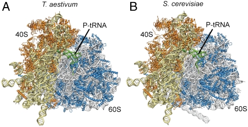
Protein synthesis in all living organisms occurs on ribonucleoprotein particles, called ribosomes. Despite the universality of this process, eukaryotic ribosomes are significantly larger in size than their bacterial counterparts due in part to the presence of 80 r proteins rather than 54 in bacteria. Using cryoelectron microscopy reconstructions of a translating plant (Triticum aestivum) 80S ribosome at 5.5-Å resolution, together with a 6.1-Å map of a translating Saccharomyces cerevisiae 80S ribosome, we have localized and modeled 74/80 (92.5%) of the ribosomal proteins, encompassing 12 archaeal/eukaryote-specific small subunit proteins as well as the complete complement of the ribosomal proteins of the eukaryotic large subunit. Near-complete atomic models of the 80S ribosome provide insights into the structure, function, and evolution of the eukaryotic translational apparatus.


Conflict of interest statement

The authors declare no conflict of interest.

Figures

Fig. 1.
Fig. 1.
Identification of r-proteins L38e and L34e. (A) Cryo-EM map of the T. aestivum 80S ribosome, with rRNA colored gray and r protein colored green. (B) Same as A, but with localized r proteins colored red. Reconstruction of (C) S. cerevisiae WT 80S ribosome compared to (D) reconstruction of S. cerevisiae 80S ribosomes isolated from a strain lacking the gene for L38e. The asterisk indicates the position of additional density assigned to L38e, and the tunnel exit (TE) is shown for reference. (E) Difference density map calculated between C and D and shown superimposed on the map from D. Reconstruction of (F) P. furiosus 70S ribosome, compared to (G) X-ray structure of the 50S subunit from H. marismortui filtered to a similar resolution. (H) Difference density map calculated between F and G and shown superimposed on the map from G identifying the location of r-protein L34e (red).
Fig. 2.
Fig. 2.
Localization of eukaryote-specific r proteins. Cryo-EM maps of the T. aestivum (A) 40S and (B) 60S subunit, with density for the newly identified r proteins colored distinctly. Molecular models of r proteins of the T. aestivum (C) 40S and (D) 60S subunit, with newly identified r proteins colored distinctly.
Fig. 3.
Fig. 3.
Functional roles for eukaryote-specific r proteins. (A) Small 40S subunit with newly modeled r-proteins S30e and S25e (red) and eukaryote-specific extension of S4p (green) highlighted (thumbnail, Left; zoom, Right). (B) Comparative view of the bacterial 30S subunit decoding site (11, 12). In A and B, the anticodon-stemloops of A-, P- and E-tRNAs (blue) and mRNA (orange) are shown for reference. (C) Large 60S subunit with eukaryote-specific extension of L10e (green) highlighted (thumbnail, Left; zoom, Right). (D) Comparative view of the bacterial 50S subunit with bacterial-specific L27p colored red (11). In C and D, the acceptor-stem of the P-tRNA (blue) is shown for reference. (E) Small 40S subunit with newly modeled r-proteins S21e, S26e, and S28e colored distinctly (thumbnail, Left; zoom, Right). (F) Comparative view of the bacterial 30S subunit with bacterial-specific S18p shown in green (11). In E and F, the P-tRNA (blue) and mRNA (orange) are shown for reference. (G and H) The binding site of eEF3 on the S. cerevisiae 80S ribosome, with (G) side and (H) top views (see insets) showing the binding site of eEF3 as a red outline and molecular models of ribosomal components that comprise the eEF3 binding site. Newly identified proteins are shown in red (S19e, S25e) and newly modeled r-protein extensions in green, whereas core r proteins are colored gray. Modified from ref. .
Fig. 4.
Fig. 4.
Coevolution of rRNA expansion segments with r proteins in the 80S ribosome. (A) Cryo-EM map of the T. aestivum 80S ribosome, with rRNA ES and variable regions colored green and eukaryote-specific r proteins and extensions colored orange. (B) View of the intertwined region of ES7L (dark blue) and ES39L (light blue), with core r proteins (gray), eukaryote-specific r-protein extensions (pale green), and r proteins (L6e, orange; L14e, red; L18ae, yellow; L28e, pink; L35ae, green) highlighted. Inset shows relative position to 40S (yellow) and 60S subunits (gray). (C) Comparison of relative positions of S4e (red) in yeast/T. aestivum 80S (Left) with S16p (green) in bacteria (11) (Right). (D) Comparison of relative positions of L29e (red) in yeast/T. aestivum 80S (Left) with L36p (green) in bacteria (11) (Right).
Fig. 5.
Fig. 5.
Structures of wheat germ and yeast eukaryotic 80S ribosomes. (A and B) Near-complete molecular models for the (A) T. aestivum and (B) S. cerevisiae 80S ribosome, with rRNA and protein shown in yellow and orange for the small subunit and gray and blue for the large subunit, respectively.

Similar articles

  • Cryo-EM structure and rRNA model of a translating eukaryotic 80S ribosome at 5.5-A resolution.
    Armache JP, Jarasch A, Anger AM, Villa E, Becker T, Bhushan S, Jossinet F, Habeck M, Dindar G, Franckenberg S, Marquez V, Mielke T, Thomm M, Berninghausen O, Beatrix B, Söding J, Westhof E, Wilson DN, Beckmann R. Armache JP, et al. Proc Natl Acad Sci U S A. 2010 Nov 16;107(46):19748-53. doi: 10.1073/pnas.1009999107. Epub 2010 Oct 27. Proc Natl Acad Sci U S A. 2010. PMID: 20980660 Free PMC article.
  • The structure and function of the eukaryotic ribosome.
    Wilson DN, Doudna Cate JH. Wilson DN, et al. Cold Spring Harb Perspect Biol. 2012 May 1;4(5):a011536. doi: 10.1101/cshperspect.a011536. Cold Spring Harb Perspect Biol. 2012. PMID: 22550233 Free PMC article. Review.
  • Structures of the human and Drosophila 80S ribosome.
    Anger AM, Armache JP, Berninghausen O, Habeck M, Subklewe M, Wilson DN, Beckmann R. Anger AM, et al. Nature. 2013 May 2;497(7447):80-5. doi: 10.1038/nature12104. Nature. 2013. PMID: 23636399
  • Eukaryotic Ribosome Assembly.
    Vanden Broeck A, Klinge S. Vanden Broeck A, et al. Annu Rev Biochem. 2024 Aug;93(1):189-210. doi: 10.1146/annurev-biochem-030222-113611. Epub 2024 Jul 2. Annu Rev Biochem. 2024. PMID: 38768392 Review.
  • Structure of the human 80S ribosome.
    Khatter H, Myasnikov AG, Natchiar SK, Klaholz BP. Khatter H, et al. Nature. 2015 Apr 30;520(7549):640-5. doi: 10.1038/nature14427. Epub 2015 Apr 22. Nature. 2015. PMID: 25901680

Cited by

References

    1. Schmeing TM, Ramakrishnan V. What recent ribosome structures have revealed about the mechanism of translation. Nature. 2009;461:1234–1242. - PubMed
    1. Sonenberg N, Hinnebusch AG. Regulation of translation initiation in eukaryotes: Mechanisms and biological targets. Cell. 2009;136:731–745. - PMC - PubMed
    1. Warner JR, McIntosh KB. How common are extraribosomal functions of ribosomal proteins? Mol Cell. 2009;34:3–11. - PMC - PubMed
    1. Freed EF, Bleichert F, Dutca LM, Baserga SJ. When ribosomes go bad: Diseases of ribosome biogenesis. Mol Biosyst. 2010;6:481–493. - PMC - PubMed
    1. Wang DO, Martin KC, Zukin RS. Spatially restricting gene expression by local translation at synapses. Trends Neurosci. 2010;33:173–182. - PMC - PubMed

Publication types

MeSH terms

LinkOut - more resources