. 2010 Nov 9;107(45):19237-41.
doi: 10.1073/pnas.1009705107. Epub 2010 Oct 25.

β-Subunit myristoylation is the gatekeeper for initiating metabolic stress sensing by AMP-activated protein kinase (AMPK)

Affiliations

β-Subunit myristoylation is the gatekeeper for initiating metabolic stress sensing by AMP-activated protein kinase (AMPK)

Jonathan S Oakhill et al. Proc Natl Acad Sci U S A. .

Abstract

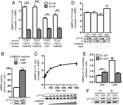
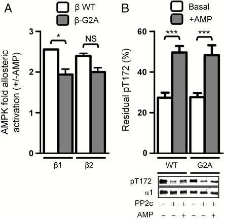
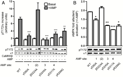
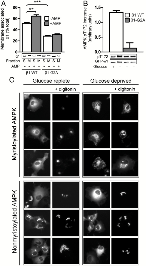
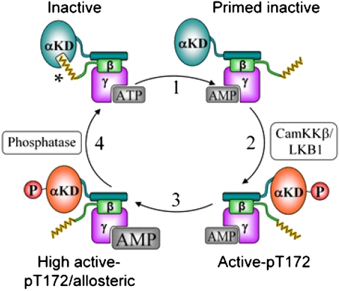
The AMP-activated protein kinase (AMPK) is an αβγ heterotrimer that acts as a master metabolic regulator to maintain cellular energy balance following increased energy demand and increases in the AMP/ATP ratio. This regulation provides dynamic control of energy metabolism, matching energy supply with demand that is essential for the function and survival of organisms. AMPK is inactive unless phosphorylated on Thr172 in the α-catalytic subunit activation loop by upstream kinases (LKB1 or calcium-calmodulin-dependent protein kinase kinase β). How a rise in AMP levels triggers AMPK α-Thr172 phosphorylation and activation is incompletely understood. Here we demonstrate unequivocally that AMP directly stimulates α-Thr172 phosphorylation provided the AMPK β-subunit is myristoylated. Loss of the myristoyl group abolishes AMP activation and reduces the extent of α-Thr172 phosphorylation. Once AMPK is phosphorylated, AMP further activates allosterically but this activation does not require β-subunit myristoylation. AMP and glucose deprivation also promote membrane association of myristoylated AMPK, indicative of a myristoyl-switch mechanism. Our results show that AMP regulates AMPK activation at the initial phosphorylation step, and that β-subunit myristoylation is important for transducing the metabolic stress signal.


Conflict of interest statement

The authors declare no conflict of interest.

Figures

Fig. 1.
Fig. 1.
Myristoylation of the β-subunit regulates AMP-activated phosphorylation of α-Thr172. In all panels, values are presented as mean ± SEM, n = 3–7. Immunoblots shown are single representative experiments or independent transfections, vertical lines indicate separate gels. (A) COS7- or E.-coli-expressed AMPK containing myristoylated (open bars) or nonmyristoylated (shaded bars) β-subunit was phosphorylated by CaMKKβ or LKB1 as indicated, in the presence or absence of AMP. The fold increase in pThr172 compared to basal, non-AMP treated controls is shown. **P < 0.01 and ***P < 0.001 compared to myristoylated AMPK. (B) COS7-expressed AMPK was phosphorylated by CaMKKβ in the presence or absence of 200 μM AMP, and AMPK activity of both was then measured in the presence of 200 μM AMP using the SAMS assay. ***P < 0.0001 compared to basally phosphorylated AMPK. (C) AMP dose response curve for the activation of CaMKKβ-mediated Thr172 phosphorylation at 120 mM NaCl. (D) AMP-activated Thr172 phosphorylation occurs with all α and β isoform combinations. (E) Myristoylation of the β-subunit inhibits basal Thr172 phosphorylation. Results shown detail CaMKKβ-mediated phosphorylation of α1β1γ1 and α1β1(G2A)γ1 from A, displayed as absolute increase in pThr172 compared to a nonphosphorylated AMPK control. **P < 0.01 compared to basally phosphorylated WT control. (F) Basal pThr172 levels of AMPK [α1β1γ1, α1β1(G2A)γ1, α1β2γ1, and α1β2(G2A)γ1] expressed in COS7 cells was measured by immunoblot after normalization for α1 expression.
Fig. 2.
Fig. 2.
Effect of AMPK β-subunit myristoylation on AMP-mediated allosteric activation and phosphatase protection. In both panels, values are presented as mean ± SEM, n = 3–7. (A) AMP-mediated allosteric activation of purified AMPK [α1β1γ1 and α1β2γ1, open bars; α1β1(G2A)γ1 and α1β2(G2A)γ1, shaded bars]. *P < 0.05; NS, not significant. (B) AMP-mediated protection against dephosphorylation of purified AMPK [α1β2γ1 and α1β2(G2A)γ1] by PP2c. Graph displays percent residual pThr172 after PP2c treatment compared to a nonphosphatase treated AMPK control. ***P < 0.001 compared to basal dephosphorylation. Immunoblot shown is a single representative experiment.
Fig. 3.
Fig. 3.
Contribution of individual γ1 AMP binding sites to AMPK regulation by AMP. In both panels, values are presented as mean ± SEM, n = 3–7. (A) WT AMPK and indicated β1/γ1 mutants were phosphorylated by CaMKKβ in the presence or absence of AMP. Graph shows the absolute increase in pThr172 compared to nonphosphorylated AMPK controls. **P < 0.01 and ***P < 0.001 compared to AMP-treated WT control. Immunoblot shown is a single representative experiment, vertical line indicates separate gels. (B) AMP-mediated allosteric activation of WT AMPK and indicated β1/γ1 mutants following CaMKKβ activation. *P < 0.05, **P < 0.01, and ***P < 0.001 compared to WT. Immunoblot shows pThr172 level of each sample assayed.
Fig. 4.
Fig. 4.
Effect of AMP and nutrient stress on myristoyl-regulated AMPK membrane association. In all panels, values are presented as mean ± SEM, n = 3–5. Immunoblots shown are single representative experiments. (A) Incubation of purified, COS7 cell-expressed AMPK [α1β1γ1 or α1β1(G2A)γ1] with liposomes (80∶20 (wt/wt) POPC∶POPS) in the presence or absence of AMP. The α1 content of soluble (S) and membrane (M) fractions were detected by immunoblot. **P < 0.01 and ***P < 0.001 compared to WT AMPK basal membrane association (see also Fig. S6). (B) Activation of AMPK in COS7 cells in response to glucose deprivation. COS7 cells expressing myristoylated or nonmyristoylated AMPK were incubated for 60 min in high or no glucose medium. Cells were harvested and pThr172 in lysates was measured by immunoblot after normalization for GFP-α1. (C) Subcellular localization of AMPK in COS7 cells in response to glucose deprivation. COS7 cells were transfected to express myristoylated (Top) or nonmyristoylated (Bottom) AMPK, and GFP-fusion α1-subunit was visualized by fluorescence microscopy under glucose replete conditions (Left) or after 60 min glucose deprivation (Right). Cells were also treated with digitonin to remove nonmembrane-bound AMPK. Images are representative of individual treatments.
Fig. 5.
Fig. 5.
Illustration of the elements of AMPK regulation by AMP. When cellular ATP levels are replete myristoyl-group (yellow) sequestration suppresses Thr172 phosphorylation and maintains the inactive state (Upper Left). Increased AMP/ATP ratio triggers a myristoyl-switch, promoting AMPK membrane association when required (step 1) and Thr172 phosphorylation (step 2). AMPK can be allosterically activated and protected from pThr172 dephosphorylation by AMP (step 3). Restoration of intracellular ATP levels reverses the myristoyl-switch (step 4). αKD, α-subunit kinase domain; * putative myristoyl binding site on α.

Similar articles

Cited by

References

    1. Hardie DG. AMP-activated/SNF1 protein kinases: Conserved guardians of cellular energy. Nat Rev Mol Cell Biol. 2007;8:774–785. - PubMed
    1. Steinberg GR, Kemp BE. AMPK in health and disease. Physiol Rev. 2009;89:1025–1078. - PubMed
    1. Crute BE, Seefeld K, Gamble J, Kemp BE, Witters LA. Functional domains of the alpha1 catalytic subunit of the AMP-activated protein kinase. J Biol Chem. 1998;273:35347–35354. - PubMed
    1. Iseli TJ, et al. AMP-activated protein kinase beta subunit tethers alpha and gamma subunits via its C-terminal sequence (186–270) J Biol Chem. 2005;280:13395–13400. - PubMed
    1. Townley R, Shapiro L. Crystal structures of the adenylate sensor from fission yeast AMP-activated protein kinase. Science. 2007;315:1726–1729. - PubMed

Publication types

LinkOut - more resources