. 2010 Nov 22;207(12):2595-607.
doi: 10.1084/jem.20100786. Epub 2010 Oct 25.

Epithelial reticulon 4B (Nogo-B) is an endogenous regulator of Th2-driven lung inflammation

Affiliations

Epithelial reticulon 4B (Nogo-B) is an endogenous regulator of Th2-driven lung inflammation

Paulette L Wright et al. J Exp Med. .

Abstract

Nogo-B is a member of the reticulon family of proteins (RTN-4B) that is highly expressed in lung tissue; however, its function remains unknown. We show that mice with Th2-driven lung inflammation results in a loss of Nogo expression in airway epithelium and smooth muscle compared with nonallergic mice, a finding which is replicated in severe human asthma. Mice lacking Nogo-A/B (Nogo-KO) display an exaggerated asthma-like phenotype, and epithelial reconstitution of Nogo-B in transgenic mice blunts Th2-mediated lung inflammation. Microarray analysis of lungs from Nogo-KO mice reveals a marked reduction in palate lung and nasal clone (PLUNC) gene expression, and the levels of PLUNC are enhanced in epithelial Nogo-B transgenic mice. Finally, transgenic expression of PLUNC into Nogo-KO mice rescues the enhanced asthmatic-like responsiveness in these KO mice. These data identify Nogo-B as a novel protective gene expressed in lung epithelia, and its expression regulates the levels of the antibacterial antiinflammatory protein PLUNC.


Figures
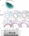
Figure 1.
Figure 1.
Nogo-B is highly expressed in the lung. (A) Whole-mount X-gal staining of Nogo-A/B KO LacZ mouse lung tissue. Blue indicates positive Nogo-A/B gene expression throughout the lobe. Bar, 0.5 mm. (B) X-gal–stained lung sections from Nogo-A/B+/− LacZ mice (top). Blue indicates positive Nogo gene expression in epithelium and smooth muscle of airways (A), and endothelial and smooth muscle cells of vessels (V), as well as parenchymal cells. Also shown are lung sections from WT mice stained with Nogo-A/B antibody 1761 (bottom). Brown staining indicates positive Nogo protein expression, and specificity of antibody was confirmed with negative staining of Nogo-A/B KO lung section (inset). Sections were also labeled with anti–E cadherin (Ecad) or α-SMA to delineate epithelium and smooth muscle, respectively. Bars: (top) 200 µm; (bottom) 100 µm. (C) Lysates were immunoblotted with a Nogo-A/B antibody. Brain lysate is as a positive control for Nogo-A. Heart and mouse lung endothelial cells (mlecs) are positive controls for Nogo-B. Lung lysate from Nogo-A/B KO mice is a negative control and hsp90 is a loading control.
Figure 2.
Figure 2.
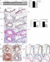
Regulation of Nogo-B expression during asthma-like conditions in mice and human asthma. (A) Nogo-B protein levels were significantly decreased in total lung lysates of OVA-sensitized and challenged (allergic) mice versus WT control (nonallergic) mice. Shown is a typical Western blot from six individual experiments. Nogo-B levels were quantified by densitometry and normalized to hsp90 (*, P < 0.05). Data are expressed as mean ± SEM. n = 4–7 mice per group. (B) Lung sections from representative small and large airways of untreated (control, a and b) and OVA-challenged (c and d) WT mice stained with Nogo-A/B antibody. Brown indicates positive Nogo-A/B in airway epithelium (arrowheads), smooth muscle and vessels (asterisk) of control mice, and the loss of Nogo immunoreactivity in epithelium and smooth muscle, but not vessels (*, c and d), of OVA-challenged mice. Bar, 100 µm. The graph on the right depicts quantification of Nogo-A/B staining with multiple sections from multiple mice using the scoring system, where 3 is 100% positive staining and 0 is an absence of staining (*, P < 0.01). Data are expressed as the mean ± SEM. n = 4–8 mice for both groups and data are representative of three experiments. Serial sections were stained with E-cadherin (e and f), H&E (g and h), and PAS (i and j). (C) Representative images of large (a and b) and small (c and d) airways from human lung sections of normal and fatal asthmatic specimens stained for Nogo with the N-18 antibody. Airway epithelium is indicated with arrowheads and vessels with asterisks. Bars: (top) 200 µm; (bottom) 100 µm. Images are representative of four individual normal human lung specimens and one individual fatal asthmatic specimen, with additional patient samples in Fig. S2. (D) Lung sections from control (top) and OVA-treated (bottom) Nogo-A/B+/− LacZ mice stained for β-gal. Airways (both E-cadherin and SMA positive) are indicated with A and vessels (SMA positive only) are indicated with V. Bars, 100 µm. Images represent results obtained from three individual experiments where n = 6 mice.
Figure 3.
Figure 3.
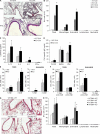
Nogo-A/B KO mice display exaggerated antigen-driven Th2 inflammatory responses. (A) Representative lung sections from allergic WT and Nogo-A/B KO (KO). (B) Total and differential cell counts in BAL fluid from WT and KO control and OVA-challenged mice (*, P < 0.01). (C) Levels of Th2 cytokines IL-13, IL-4, and IL-5 and IFN-γ as a Th1 cytokine for control were measured in BAL of WT and KO OVA mice (*, P < 0.05). (D) Levels of OVA-specific IgE in serum of WT and KO control mice and in OVA mice before (sensitized only) and after challenge (*, P = 0.013). (E) Total cells and eosinophils were measured in BAL of allergic WT and KO mice as described in Results. *, statistically significant from WT mice. (F) 7 and 14 d after 3-d challenge (*, P < 0.05). (G and H) Lung histology (G) and cell counts (H) in BAL in OVA-challenged BMT mice (*, P < 0.05). Data are expressed as the mean ± SEM. n = 4–8 mice for all groups representative of four individual experiments. Bars, 100 µm.
Figure 4.
Figure 4.
Transgenic expression of Nogo-B in lung epithelium inhibits allergic Th2 inflammation. (A) CCSP-Nogo-DTG mice were put on doxycycline water (0.5 mg/ml) for different time points and the levels of Nogo-B HA–tagged transgene assessed. Endogenous mouse Nogo-B and hsp90 levels were used as loading controls. Each lane represents lung lysate from an individual mouse and was repeated one additional time. (B) Transgene expression in lung sections from CCSP-Ng-DTG mice. Bars, 100 µm. (C) CCSP-Ng-STG and DTG littermates were put on doxycycline water for 7 d before OVA sensitization/challenge. Lung sections from control and OVA-sensitized and challenged STG and DTG mice were stained for transgene expression (a and b, and c and d, respectively), Nogo-A/B (e and f), H&E (g and h), and PAS (I and j). Arrowheads highlight airway epithelium. Bar, 200 µm. (D and E) BAL cell counts (D) and levels of cytokines (E) were evaluated from BAL of OVA STG and DTG mice (*, P < 0.05). (F) WT OTII CD4+ Th2 cells were purified and adoptively transferred into STG and DTG mice, followed by OVA challenge and evaluation of total levels of inflammation in the BAL (*, P < 0.05). Data are expressed as the mean ± SEM. n = 5–8 mice for both groups representative of three individual experiments.
Figure 5.
Figure 5.
Nogo regulates Plunc expression in vivo and in vitro. (A) Levels of basal gene expression (PLUNC, Tff2, Reg3γ, IL13Rα1, IL13Rα2, and amcase) in WT and Nogo-A/B KO (KO) lungs were determined by qRT-PCR. Data were expressed relative to WT gene expression of 100% (*, P < 0.01). (B) PLUNC gene expression in lungs from control and allergic mice. Data are expressed relative to WT control gene expression of 100% (*, P < 0.01). (C and D) Basal levels of PLUNC protein were analyzed in BAL of WT and Nogo-A/B−/− mice (C) and in BAL of CCSP-Ng-STG and –DTG mice (D) put on doxycycline water for 3 mo using a PLUNC antibody. Ponceau-stained band at 150 Kb was used as a loading control. (E) NHBECs were treated with a nonsilencing (NS) siRNA or siRNA specifically targeting Nogo-B for 72 h and evaluated for gene expression of Nogo-B, Plunc, TGF-β, and IL4Rα by qRT-PCR. Data were expressed relative to NS gene expression of 100% (*, P < 0.01). Data are expressed as the mean ± SEM. n = 3–6 mice for all groups representative of three experiments with each sample run in triplicate.
Figure 6.
Figure 6.
Transgenic expression of PLUNC in lungs of Nogo-A/B KO mice reduces allergic Th2 inflammation. (A) Lung sections from WT, KO, PLUNC transgenic (PTG)–WT, and PTG-KO were stained for PLUNC. Endogenous PLUNC (only in the trachea and upper airways) was observed in tracheal epithelium of WT and, to a lesser extent, in KO (top). Transgenic PLUNC was observed in epithelium of upper airways (bottom) as well as further down the smaller airways (insets) in both WT and KO backgrounds. (B) BAL from WT, PTG-WT, KO, and PTG-KO mice with and without OVA challenge were immunoblotted for levels of endogenous PLUNC (mPLUNC at 25 kD) and transgenic PLUNC (hPLUNC at 20 kD) protein expression. Each lane of the Western blot represents lung lysate from typical individual mouse and was observed in n = 4–8 mice for each group. (C and D) Lung H&E staining (C) and total and differential BAL cell counts (D) of KO and PTG-KO mice challenged with OVA (*, P < 0.05; **, P < 0.01). Bars, 200 µm. Data are expressed as the mean ± SEM. n = 6 mice for both groups representative of two experiments.

Similar articles

Cited by

References

    1. Acevedo L., Yu J., Erdjument-Bromage H., Miao R.Q., Kim J.E., Fulton D., Tempst P., Strittmatter S.M., Sessa W.C. 2004. A new role for Nogo as a regulator of vascular remodeling. Nat. Med. 10:382–388 10.1038/nm1020 - DOI - PubMed
    1. Bingle C.D., LeClair E.E., Havard S., Bingle L., Gillingham P., Craven C.J. 2004. Phylogenetic and evolutionary analysis of the PLUNC gene family. Protein Sci. 13:422–430 10.1110/ps.03332704 - DOI - PMC - PubMed
    1. Cantero-Recasens G., Fandos C., Rubio-Moscardo F., Valverde M.A., Vicente R. 2010. The asthma-associated ORMDL3 gene product regulates endoplasmic reticulum-mediated calcium signaling and cellular stress. Hum. Mol. Genet. 19:111–121 10.1093/hmg/ddp471 - DOI - PubMed
    1. Chen M.S., Huber A.B., van der Haar M.E., Frank M., Schnell L., Spillmann A.A., Christ F., Schwab M.E. 2000. Nogo-A is a myelin-associated neurite outgrowth inhibitor and an antigen for monoclonal antibody IN-1. Nature. 403:434–439 10.1038/35000219 - DOI - PubMed
    1. Chu H.W., Thaikoottathil J., Rino J.G., Zhang G., Wu Q., Moss T., Refaeli Y., Bowler R., Wenzel S.E., Chen Z., et al. 2007. Function and regulation of SPLUNC1 protein in Mycoplasma infection and allergic inflammation. J. Immunol. 179:3995–4002 - PubMed

Publication types