. 2010 Jun 15;1(3):27.
doi: 10.1038/ncomms1027.

Imaging the nanoscale organization of peptidoglycan in living Lactococcus lactis cells

Affiliations
Free PMC article

Imaging the nanoscale organization of peptidoglycan in living Lactococcus lactis cells

Guillaume Andre et al. Nat Commun. .
Free PMC article

Abstract

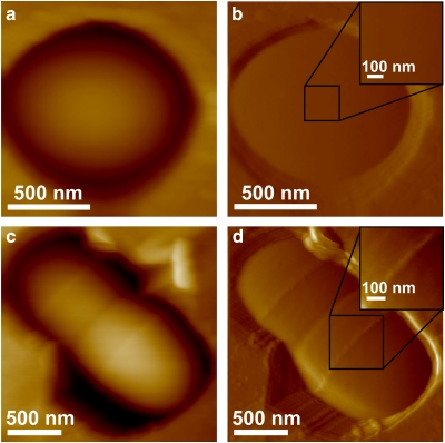
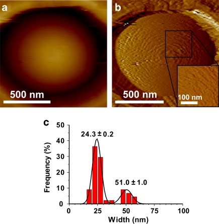
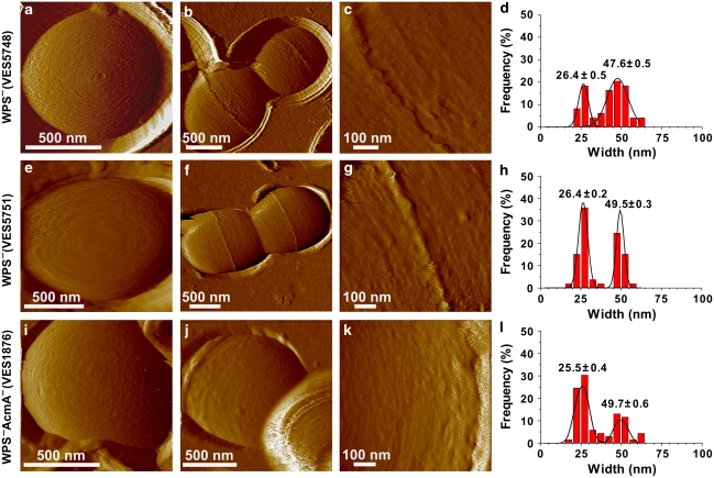
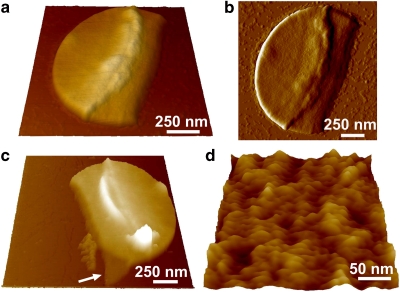
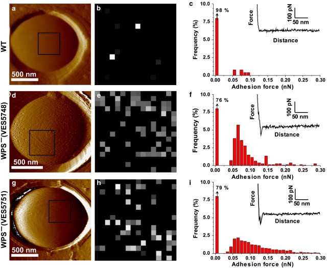
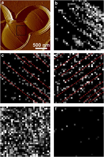
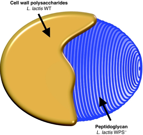
The spatial organization of peptidoglycan, the major constituent of bacterial cell-walls, is an important, yet still unsolved issue in microbiology. In this paper, we show that the combined use of atomic force microscopy and cell wall mutants is a powerful platform for probing the nanoscale architecture of cell wall peptidoglycan in living Gram-positive bacteria. Using topographic imaging, we found that Lactococcus lactis wild-type cells display a smooth, featureless surface morphology, whereas mutant strains lacking cell wall exopolysaccharides feature 25-nm-wide periodic bands running parallel to the short axis of the cell. In addition, we used single-molecule recognition imaging to show that parallel bands are made of peptidoglycan. Our data, obtained for the first time on living ovococci, argue for an architectural feature of the cell wall in the plane perpendicular to the long axis of the cell. The non-invasive live cell experiments presented here open new avenues for understanding the architecture and assembly of peptidoglycan in Gram-positive bacteria.


Figures

Figure 1
Figure 1. AFM images of WT L. lactis cells.
(a, c) Height and (b, d) deflection images recorded in sodium acetate buffer for L. lactis WT cells using a small applied force (250 pN). The images show either the pole of a single cell (a, b) or two cells during the course of the division process (c, d). Insets in b and d show high-resolution deflection images recorded in the square regions.
Figure 2
Figure 2. Imaging WT cells at large forces causes substantial ultrastructural changes.
(a) Height and (b) deflection images of the polar region of an L. lactis WT cell recorded with an applied force of 5 nN. The inset shows a high-resolution deflection image recorded in the square region. (c) Statistical analysis of the width of the periodic features observed in height images (n=45 measurements on two different cells, average width ± s.e.m.).
Figure 3
Figure 3. AFM images of mutant strains reveal periodic bands running parallel to the short cell axis.
Deflection images of polar regions (a, e, i) and of dividing bacteria (b, c, f, g, j, k) recorded for L. lactis VES5748 (ac) and VES5751 (eg) WPS mutants lacking cell wall exopolysaccharides, and for the double mutant VES1876 WPSAcmA lacking both exopolysaccharides and the major autolysin AcmA (ik). (c, g, k) High-resolution images obtained on the longitudinal region of the cells. All images were recorded in sodium acetate buffer using a small applied force (250 pN). (d, h, i) Statistical analysis of the width of the periodic features observed in the height images of VES5748 WPS mutant (d, n=55 measurements, on five different cells), of VES5751 WPS mutant (h, n=53 measurements, on four different cells, average width ± s.e.m.) and of VES1876 WPSAcmA double mutant (i, n=57 measurements, on three different cells, average width ± s.e.m.).
Figure 4
Figure 4. Sacculi do not show parallel bands.
(a, c, d) Height and (b) deflection images in air of peptidoglycan sacculi from L. lactis VES5748, gently broken by a french press. Images a and b show the outside surface of the same sacculus consisting of a double cell wall. Image c represents another sacculus in which the boarder exposes the inner surface of a single wall (arrow). (d) High-resolution view of the outer surface, similar morphology being observed on the inner surface.
Figure 5
Figure 5. Single-molecule recognition imaging of peptidoglycan.
(a, d, g) Deflection images recorded with silicon nitride tips on L. lactis WT (a), VES5748 WPS mutant (d) and VES5751 WPS mutant (g). (b, e, h) Adhesion force maps (400×400 nm) recorded on the three strains with LysM tips in the square areas shown in the deflection images, using a maximum applied force of 250 pN. (c, f, i) Adhesion histograms generated from four adhesion force maps (n=1,024 curves), together with representative force curves.
Figure 6
Figure 6. Peptidoglycan localizes as parallel lines on the surface of WPS mutants.
(a) Deflection image of an L. lactis VES5748 WPS mutant cell recorded with a silicon nitride tip. (b) Adhesion force map (400×400 nm) recorded with an LysM tip in the square area shown in the deflection image using a maximum applied force of 250 pN. (cf) Adhesion force maps (500×500 nm) recorded with an LysM tip on another cell. Maps shown in c and d were recorded in the same area, except that the cell was rotated by 90°. Many of the detected molecules (bright pixels) were arranged as lines running parallel to the short cell axis (red lines). The map shown in e was obtained with a maximum applied force of 500 pN instead of 250 pN, whereas the map in f was obtained in a 10 μg ml−1 peptidoglycan solution. In addition to the data shown, similar results were obtained in nine different cells from at least five different cultures, using 10 different tips from at least five different batches.
Figure 7
Figure 7. Schematic drawing of the nanoscale organization of the L. lactis peptidoglycan.
The cartoon emphasizes the outermost surface layers, that is, cell wall polysaccharides in WT cells, and peptidoglycan arranged as periodic bands in WPS cells.

References

    1. Beveridge T. J. & Graham L. L. Surface layers of bacteria. Microbiol. Mol. Biol. Rev. 55 684–705 (1991). - PMC - PubMed
    1. Sleytr U. B. & Beveridge T. J. Bacterial S-layers. Trends Microbiol. 7 253–260 (1999). - PubMed
    1. Archibald A. R., Hancock I. C. & Harwood C. R. in Bacillus Subtilis and Other Gram-Positive Bacteria: Biochemistry, Physiology, and Molecular Genetics (Sonenshein, A. L., Hoch, J. A. & Losick, R., eds) 381–410 (American Society for Microbiology, 1993).
    1. Costerton J., Ingram J. M. & Cheng K. J. Structure and function of cell-envelope of Gram-negative bacteria. Bacteriol. Rev. 38 87–110 (1974). - PMC - PubMed
    1. Delcour J., Ferain T., Deghorain M., Palumbo E. & Hols P. The biosynthesis and functionality of the cell-wall of lactic acid bacteria. Anton Leeuw Int. J. G 76 159–184 (1999). - PubMed

Publication types

MeSH terms