. 2010 Oct 21;6(10):e1001170.
doi: 10.1371/journal.pgen.1001170.

Dual functions of ASCIZ in the DNA base damage response and pulmonary organogenesis

Affiliations

Dual functions of ASCIZ in the DNA base damage response and pulmonary organogenesis

Sabine Jurado et al. PLoS Genet. .

Erratum in

Abstract

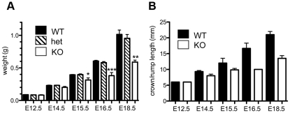
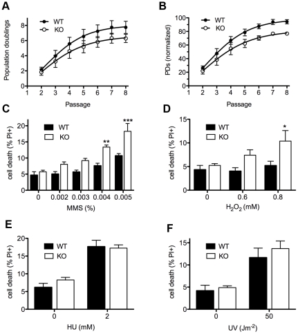
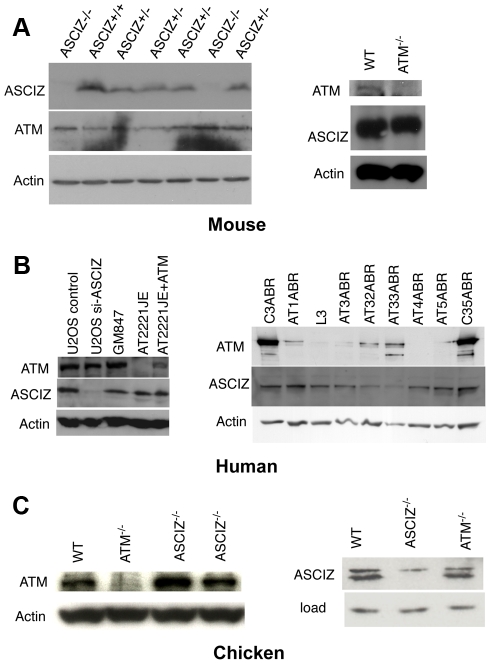
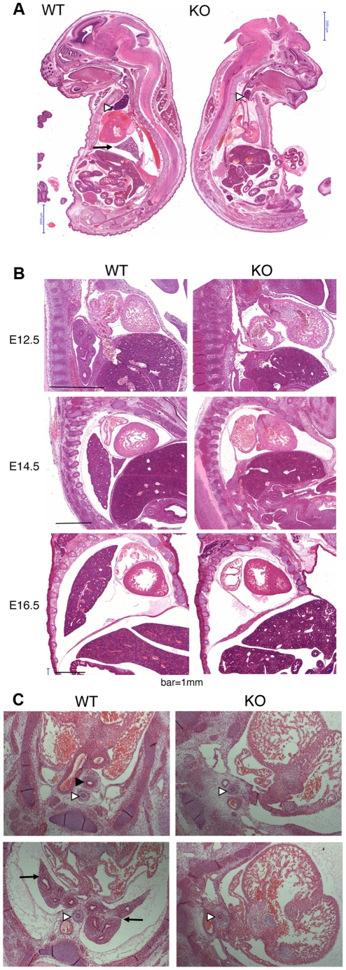
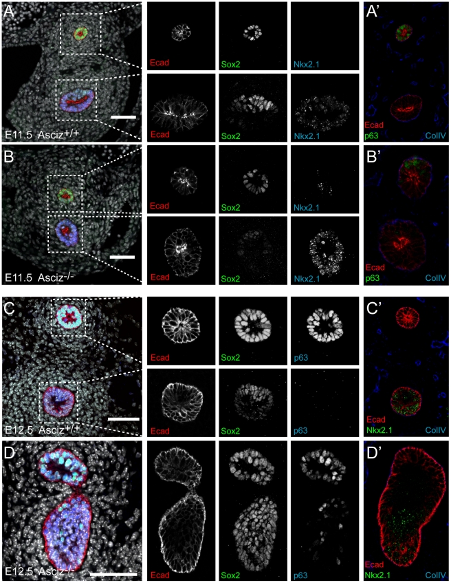
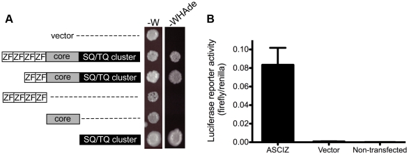
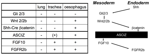
Zn²(+)-finger proteins comprise one of the largest protein superfamilies with diverse biological functions. The ATM substrate Chk2-interacting Zn²(+)-finger protein (ASCIZ; also known as ATMIN and ZNF822) was originally linked to functions in the DNA base damage response and has also been proposed to be an essential cofactor of the ATM kinase. Here we show that absence of ASCIZ leads to p53-independent late-embryonic lethality in mice. Asciz-deficient primary fibroblasts exhibit increased sensitivity to DNA base damaging agents MMS and H2O2, but Asciz deletion knock-down does not affect ATM levels and activation in mouse, chicken, or human cells. Unexpectedly, Asciz-deficient embryos also exhibit severe respiratory tract defects with complete pulmonary agenesis and severe tracheal atresia. Nkx2.1-expressing respiratory precursors are still specified in the absence of ASCIZ, but fail to segregate properly within the ventral foregut, and as a consequence lung buds never form and separation of the trachea from the oesophagus stalls early. Comparison of phenotypes suggests that ASCIZ functions between Wnt2-2b/ß-catenin and FGF10/FGF-receptor 2b signaling pathways in the mesodermal/endodermal crosstalk regulating early respiratory development. We also find that ASCIZ can activate expression of reporter genes via its SQ/TQ-cluster domain in vitro, suggesting that it may exert its developmental functions as a transcription factor. Altogether, the data indicate that, in addition to its role in the DNA base damage response, ASCIZ has separate developmental functions as an essential regulator of respiratory organogenesis.


Conflict of interest statement

The authors have declared that no competing interests exist.

Figures

Figure 1
Figure 1. Generation of Asciz-deficient mice.
(A) Schematic comparison of human and mouse ASCIZ. ZF, Zn2+ finger; NLS, nuclear localization signal. Lollipops indicate predicted ATM/ATR phosphorylation sites. (B) Asciz gene structure and targeting strategy, drawn approximately to scale. The four exons (A–D) are indicated by black boxes, as are locations of oligonucleotide primers, ScaI restriction sites and the probe for genotyping, and the positions of loxP sites. (C) Southern blot (top) and PCR genotyping (bottom) of a randomly chosen litter from a heterozygote intercross at weaning. (D) PCR genotyping of a randomly chosen litter at E15.5. (E) Western blot analysis of head extracts of a randomly chosen litter at E12.5 using the indicated antibodies. (F) Western blot analysis of the indicated tissues of an 8-week old male WT mouse, and E15.5 Asciz+/− and Asciz−/− head extracts as antibody specificity controls.
Figure 2
Figure 2. Late gestational growth defect of Asciz-deficient embryos.
(A) Embryo weights of WT, heterozygotes and Asciz-deficient embryos at the indicated times post-conception. Data are the mean ± standard error; 22–55 embryos were analysed per timepoint (KOs: n = 8 (E12.5), 12 (E14.5), 8 (E15.5), 11 (E16.5), and 4 (E18.5). (B) Crown-rump length of embryos determined by histomorphometry. Data are mean ± standard error, n = 3–9 per data point. *p<0.05, **p<0.01, ***p<0.001.
Figure 3
Figure 3. Cellular phenotypes of Asciz-deficient primary fibroblasts.
(A) Cumulative population doublings (PDs) of Asciz-deficient and matched WT littermate MEFs in a standardized 3T3 assay. Data are mean ± standard error, n = 6. (B) Data from panel A normalized to the PD maximum in the relevant litter. (C–F) DNA damage sensitivity assays. Cells were treated with the indicated doses of MMS, H2O2, 2 mM HU or 50 J/m2 UV, and viability was determined by propidium iodide exclusion and FACS. *p<0.05, **p<0.01, ***p<0.001.
Figure 4
Figure 4. Reciprocal independence of ASCIZ and ATM protein levels.
(A) Protein levels in mouse tissues. Left panel, Western blot analysis of head extracts of a randomly chosen litter from an Asciz heterozygote intercross at E12.5. Right panel, brain extracts of WT and Atm-null littermate mice . (B) Protein levels in human cell lines. Left panel, adherent cells: U2OS osteosarcoma cells treated with GL2 control or Asciz siRNA; GM847 control fibroblasts, Atm-deficient AT2221JE fibroblasts containing an empty-vector control (FTY pEBS7) or reconstituted with WT Atm (FTYZ5) . Right panel, lymphoblastoid cell lines from healthy donors (C3ABR, C35ABR) and seven separate AT patients (L3 and AT1ABR–AT33ABR); note that ATM was immunoprecipitated before blotting as described . (C) Protein levels in chicken DT40 B cell lysates. Left panel, comparison of ATM levels in two independent Asciz-deleted clones using the anti-chicken ATM antibody and the ATM-deleted DT40 clone as specificity control. Right panel, comparison of ASCIZ levels in WT and an Atm-deleted clone with an Asciz-deficient clone as antibody specificity control (NB, anti-human ASCIZ was used at 1∶100 dilution rather than 1∶2000–1∶4000 for mouse or human samples).
Figure 5
Figure 5. Histological analysis of Asciz-null embryos.
(A) Sagittal sections of comparable levels of WT and Asciz−/− littermates at E18.5. Note the absence of lung (arrow), hypoplastic thymus (arrowhead), compressed thorax, steep ascending aorta, and exencephaly in the Asciz-null embryo. This embryo also represents an isolated case of omphalocele. Scale bars = 2 mm. (B) Micrographs of comparable sagittal sections of WT and Asciz−/− littermates at E12.5–16.5. Note the apparent caudal drop of the atrium relative to the ventricle in Asciz null embryos compared to WT littermates where the atrium seems to be propped up by the developing left lung. Scale bars = 1 mm. (C) Micrographs of comparable transverse sections of E12.5 WT and Asciz−/− littermates at the upper (top panels) and lower levels (bottom panels) of the thorax. Open arrowheads point to the oesophagus, the filled arrowhead and arrows point at the trachea and lungs respectively that are only present in the WT.
Figure 6
Figure 6. Defective pulmonary and tracheal development in Asciz-null embryos.
Optical projection tomography of whole-mount E-cadherin stained of E11.5 (A, B) and E12.5 (C, D) littermates. Stippled boxes indicate the approximate plane of sections chosen for immunofluorescence analysis in Figure 7. Panels are arranged with the oesophagus on top.
Figure 7
Figure 7. Expression analysis of markers of foregut development.
Sections from the levels indicated in Figure 6 were stained with the indicated antibodies. All panels are oriented with the oesophagus or dorsal foregut on top. A′–D′ are sections adjacent to the ones shown in A–D. In the merged panel on the left, nuclei are counterstained with DAPI. Scale bars = 20 µm.
Figure 8
Figure 8. ASCIZ has transcription activating function in reporter assays.
(A) Yeast one-hybrid assay. Yeast strains containing the empty vector expressing the Gal4-DBD only or the indicated human ASCIZ constructs (“long form”, residues 1–823; “short form”, residues 156–823; ZnF domain, residues 67–223; “core domain”, residues 230–442; SQ/TQ cluster domain, residues 432–823) were spotted onto -W plates as a loading control and -WHAde plates as an assay for activation of the GAL1-HIS3 and GAL2-ADE2 reporter genes. (B) Dual luciferase reporter assay of human U2OS cells transfected with pCDNA3-Gal4DBD or Gal4DBD-ASCIZ667.
Figure 9
Figure 9. Comparison of the Asciz−/− phenotype to other mouse mutants with pulmonary agenesis.
Table summarizing comparable phenotypes and schematic diagram of their role in the crosstalk between endodermal and mesodermal signaling pathways regulating early respiratory tract development; see discussion for details.

References

    1. Jackson SP, Bartek J. The DNA-damage response in human biology and disease. Nature. 2009;461:1071–1078. - PMC - PubMed
    1. Shiloh Y. ATM and related protein kinases: safeguarding genome integrity. Nat Rev Cancer. 2003;3:155–168. - PubMed
    1. Barnes DE, Lindahl T. Repair and genetic consequences of endogenous DNA base damage in mammalian cells. Annu Rev Genet. 2004;38:445–476. - PubMed
    1. Almeida KH, Sobol RW. A unified view of base excision repair: lesion-dependent protein complexes regulated by post-translational modification. DNA Repair (Amst) 2007;6:695–711. - PMC - PubMed
    1. Horton JK, Wilson SH. Hypersensitivity phenotypes associated with genetic and synthetic inhibitor-induced base excision repair deficiency. DNA Repair (Amst) 2007;6:530–543. - PMC - PubMed

Publication types

MeSH terms