. 2010 Oct 19;8(10):e1000520.
doi: 10.1371/journal.pbio.1000520.

Frequent and efficient use of the sister chromatid for DNA double-strand break repair during budding yeast meiosis

Affiliations

Frequent and efficient use of the sister chromatid for DNA double-strand break repair during budding yeast meiosis

Tamara Goldfarb et al. PLoS Biol. .

Abstract

Recombination between homologous chromosomes of different parental origin (homologs) is necessary for their accurate segregation during meiosis. It has been suggested that meiotic inter-homolog recombination is promoted by a barrier to inter-sister-chromatid recombination, imposed by meiosis-specific components of the chromosome axis. Consistent with this, measures of Holliday junction-containing recombination intermediates (joint molecules [JMs]) show a strong bias towards inter-homolog and against inter-sister JMs. However, recombination between sister chromatids also has an important role in meiosis. The genomes of diploid organisms in natural populations are highly polymorphic for insertions and deletions, and meiotic double-strand breaks (DSBs) that form within such polymorphic regions must be repaired by inter-sister recombination. Efforts to study inter-sister recombination during meiosis, in particular to determine recombination frequencies and mechanisms, have been constrained by the inability to monitor the products of inter-sister recombination. We present here molecular-level studies of inter-sister recombination during budding yeast meiosis. We examined events initiated by DSBs in regions that lack corresponding sequences on the homolog, and show that these DSBs are efficiently repaired by inter-sister recombination. This occurs with the same timing as inter-homolog recombination, but with reduced (2- to 3-fold) yields of JMs. Loss of the meiotic-chromosome-axis-associated kinase Mek1 accelerates inter-sister DSB repair and markedly increases inter-sister JM frequencies. Furthermore, inter-sister JMs formed in mek1Δ mutants are preferentially lost, while inter-homolog JMs are maintained. These findings indicate that inter-sister recombination occurs frequently during budding yeast meiosis, with the possibility that up to one-third of all recombination events occur between sister chromatids. We suggest that a Mek1-dependent reduction in the rate of inter-sister repair, combined with the destabilization of inter-sister JMs, promotes inter-homolog recombination while retaining the capacity for inter-sister recombination when inter-homolog recombination is not possible.


Conflict of interest statement

The authors have declared that no competing interests exist.

Figures

Figure 1
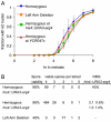
Figure 1. Similar timing of IS and IH DSB repair.
(A) Structure of DSB hotspots in a 3.5-kb his4::URA3-arg4 insert and in the YCR047c–YCR048w intergenic region . White boxes indicate URA3-arg4 insert genes; grey boxes indicate other genes on chromosome III; horizontal bars indicate sequences used for probes ,; vertical lines indicate restriction sites used to detect DSBs (short lines) and JM intermediates (long lines). (B) Strains used. (i) Homozygous control—his4::URA3-arg4 and YCR047c–YCR048w are present on both homologs. (ii) Hemizygous at his4::URA3-arg4—the 3.5-kb his4::URA3-arg4 insert is present on only one homolog. (iii) Left arm deletion—the his4::URA3-arg4 insert is present on one homolog, opposite a 90-kb deletion on the other homolog. (iv) Hemizygous at YCR047c—4 kb of DNA between YCR046c and YCR051w is deleted from one homolog. (C) Southern blots showing detection of DSBs in wild-type and rad50S strains hemizygous for his4::URA3-arg4 at either the his4::URA3-arg4 site (left) or the YCR047c homozygous control site (right). P, parental band; DSB, DSB band. (D) DSB frequencies (3–4 independent experiments, error bars indicate SEM), quantified as percent of total lane signal. Symbols connected by lines are noncumulative DSB frequencies from RAD50 strains (left-hand y-axis); unconnected symbols at 7 h are cumulative DSB frequencies from rad50S strains are (right-hand y-axis). (E) DSB life span, calculated using 7-h rad50S cumulative DSB levels, as described previously . Underlined life spans are for DSBs that must be repaired by IS recombination. Strains in (C–E) are (for RAD50 and rad50S, respectively): homozygous, MJL3201 and MJL3198; hemizygous at his4::URA3-arg4, MJL3250 and MJL3338; left arm deletion, MJL3227 and MJL3233; and hemizygous at YCR047c, MJL3399 and MJL3408.
Figure 2
Figure 2. DSBs at a hemizygous locus do not alter nuclear division timing or spore viability.
(A) Timing of the meiosis I nuclear division, monitored by DAPI staining (see Protocol S1). Values are the average of 3–4 experiments (error bars indicate standard deviation). RAD50 strains as in Figure 1. (B) Spore viability in tetrads in the indicated strains. NMS indicates non-Mendelian segregation (full conversion and post-meiotic segregation) at the his4::URA3-arg4 insert. The strain homozygous for the insert (MJL3195) is a his4::URA3-arg4-pal/his4::ura3-pal-ARG4 trans-heterozygote. Non-Mendelian segregation at ura3-pal and at arg4-pal were scored; non-Mendelian segregation for both markers in the same tetrad was scored as a single event. In the his4::URA3-arg4 hemizygote (MJL3192), non-Mendelian segregation events involved loss (1∶3) or gain (3∶1) of the URA3-arg4 insert.
Figure 3
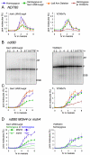
Figure 3. Reduced JM formation during IS chromatid recombination.
(A) Noncumulative JM levels in wild-type strains at the his4::URA3-arg4 insert and at YCR047c, expressed as percent of total signal in the lane. Each line represents the average of 3–4 experiments (error bars indicate SEM). (B) Southern blots of DNA from ndt80Δ strains hemizygous for his4::URA3-arg4 (MJL3497) or homozygous for his4::URA3-arg4 (MJL3252, denoted by asterisks) detecting JM intermediates at either his4::URA3-arg4 (left) or at the homozygous YCR047c site (right). P, parental. (C) JM frequencies in ndt80Δ strains, quantified as percent of total lane signal. Each point represents the average of 2–4 experiments (error bars indicate SEM). Strains used: fully homozygous, MJL3252, blue diamonds; hemizygous at his4::URA3-arg4, MJL3497, green squares; 90-kb left arm deletion, MJL3245, orange triangles; hemizygous at YCR047c–YCR048w, MJL3406, red Xs. (D) IS JM formation is Msh4-dependent. JM frequencies (error bars indicate SEM), quantified as percent of total lane signal, in DNA from ndt80Δ strains hemizygous (green) or homozygous (blue) for his4::URA3-arg4 and either MSH4 (MJL3497 or MJL3252, filled symbols) or msh4Δ (MJL3385 or MJL3386, open symbols). Each point represents the average of 2–4 experiments (error bars indicate SEM).
Figure 4
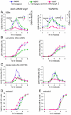
Figure 4. Altered DSB and JM metabolism in mek1Δ strains.
(A) DSB frequencies (3–4 independent experiments, error bars indicate SEM), quantified as percent of total lane signal. Symbols: blue diamonds, fully homozygous MEK1 strain (MJL3201 and MJL3198); green squares, MEK1 strains hemizygous for his4::URA3-arg4 (MJL3250 and MJL3338); and pink circles, mek1Δ strain hemizygous for his4::URA3-arg4 (MJL3370 and MJL3381). Symbols connected by lines are noncumulative DSB frequencies from RAD50 strains (left-hand y-axis; MJL3201, MJL3250, and MJL3370); unconnected symbols at 7 h are cumulative DSB frequencies from rad50S strains (right-hand y-axis; MJL3198, MJL3338, and MJL3381). (B) Cumulative JM frequencies (3–4 independent experiments, error bars indicate SEM) from ndt80Δ MEK1 and ndt80Δ mek1Δ strains. Symbols: blue diamonds, fully homozygous MEK1 strain (MJL3252); green squares, MEK1 strains hemizygous for his4::URA3-arg4 (MJL3497); and pink circles, mek1Δ strain hemizygous for his4::URA3-arg4 (MJL3387). (C) Noncumulative JM frequencies (3–4 independent experiments, error bars indicate SEM) from the same RAD50 strains used for DSB analysis in (A). (D and E) Timing of the meiosis I (D) and meiosis II (E) nuclear divisions, monitored by DAPI staining (see Protocol S1; 3–4 independent experiments, error bars indicate standard deviation), in the same RAD50 strains used for DSB and JM analysis in (A) and (C).
Figure 5
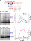
Figure 5. IS JM formation in mek1Δ.
(A) Recombination assay system used to distinguish IS and IH JMs. The URA3-arg4 construct is inserted at LEU2 on one homolog (red) and at HIS4 on the other homolog (blue). Digestion with XmnI (X) produces IS JMs and IH JMs that can be distinguished on the basis of electrophoretic mobility. (B) Representative Southern blot of DNA from a mek1Δ ndt80Δ strain with his4::URA3-arg4 and leu2::URA3-arg4 insert (MJL3397). (C) Frequencies (left y-axis, percent of total lane signal, three independent experiments, error bars indicate SEM) of IH JMs (pink squares, his4-leu2 band in [B]) and IS JMs (green diamonds, sum of his4-his4 and leu2-leu2 bands in [B]). Grey circles: IS/IH JM ratio (right y-axis). (D) Representative Southern blot of DNA from a MEK1 ndt80Δ strain with his4::URA3-arg4 and leu2::URA3-arg4 insert (MJL3523). (E) Frequencies (left y-axis, percent of total lane signal, two independent experiments, error bars indicate SEM) of IH JMs (pink squares, his4-leu2 band in [D]) and IS JMs (green diamonds, sum of his4-his4 and leu2-leu2 bands in [D]). Grey circles: IS/IH JM ratio (right y-axis).
Figure 6
Figure 6
(A) Timing of molecular events at a hemizygous (MJL3250) and homozygous (MJL3201) his4::URA3-arg4 insert. Left- and right-hand edges of rectangles indicate half-maximum points on cumulative curves for formation and repair/resolution, respectively. For meiotic divisions, left- and right-hand edges indicate 50% times for meiosis I (M1) and meiosis II (M2), respectively. Times are normalized by setting the 50% time for meiosis II to 6 h (actual times ± standard deviation: MJL3201, 5.66±0.27 h; MJL3250, 6.08±0.33 h). Left- and right-hand error bars denote the standard deviation for the half-maximum value and the sum of the standard deviations of the life span and the half-maximum value, respectively. (B) Estimation of IS/IH ratio for all recombination events at homozygous loci in wild-type, based on the following: (1) about 1/2 of IH events involve JMs; (2) about 1/4 to 1/6 of IS events involve JMs; (3) about 1/5 of JMs are IS (numbers in parentheses are observed range in the literature). Based on these values, an IS/IH total event ratio of 1∶1.7 to 1∶2.5 is calculated. Detailed calculations and IS/IH total event ratios for the full range of IS/IH JM ratios are in Protocol S1 and Figure S2. (C) How localized kinase activation can cause selective retardation of IS recombination. DSBs form when potential DSB sites on cohesed sister chromatids (pink boxes) are recruited to the chromosome axis (green). Mec1/Tel1 checkpoint kinases are activated by DSBs and associated single-stranded DNA, and phosphorylate chromatin and axis proteins in the vicinity of DSBs (red). Phosphorylated axis proteins recruit and activate Mek1 kinase, which phosphorylates target proteins (including strand transferase accessory proteins) in the vicinity of the DSB-activated axis. Strand invasion of the sister chromatid, which is within the zone of axis-associated inhibition, is thus kinetically impeded; strand invasion of the homolog is unaffected.

References

    1. Hassold T, Hall H, Hunt P. The origin of human aneuploidy: where we have been, where we are going. Hum Mol Genet. 2007;16 Spec No. 2:R203–R208. - PubMed
    1. Keeney S, Giroux C. N, Kleckner N. Meiosis-specific DNA double-strand breaks are catalyzed by Spo11, a member of a widely conserved protein family. Cell. 1997;88:375–384. - PubMed
    1. San Filippo J, Sung P, Klein H. Mechanism of eukaryotic homologous recombination. Annu Rev Biochem. 2008;77:229–257. - PubMed
    1. Kadyk L. C, Hartwell L. H. Sister chromatids are preferred over homologs as substrates for recombinational repair in Saccharomyces cerevisiae. Genetics. 1992;132:387–402. - PMC - PubMed
    1. Bzymek M, Thayer N. H, Oh S. D, Kleckner N, Hunter N. Double Holliday junctions are intermediates of DNA break repair. Nature. 2010;464:937–941. - PMC - PubMed

Publication types

MeSH terms

Substances