. 2010 Oct 18;5(10):e13453.
doi: 10.1371/journal.pone.0013453.

Reversible block of mouse neural stem cell differentiation in the absence of dicer and microRNAs

Affiliations

Reversible block of mouse neural stem cell differentiation in the absence of dicer and microRNAs

Therese Andersson et al. PLoS One. .

Abstract

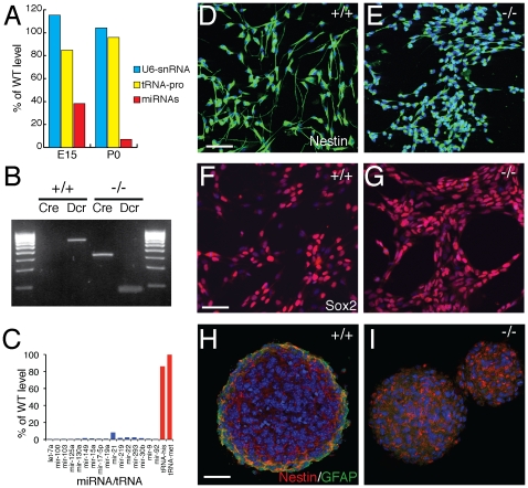
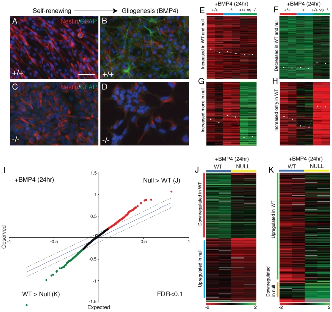
Background: To investigate the functions of Dicer and microRNAs in neural stem (NS) cell self-renewal and neurogenesis, we established neural stem cell lines from the embryonic mouse Dicer-null cerebral cortex, producing neural stem cell lines that lacked all microRNAs.

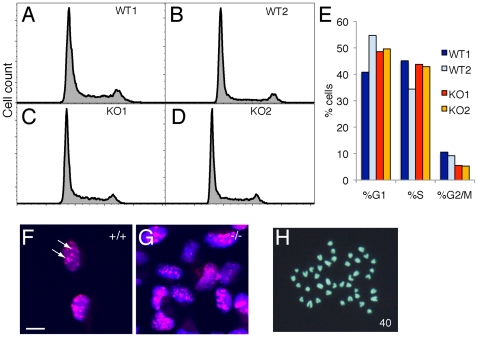
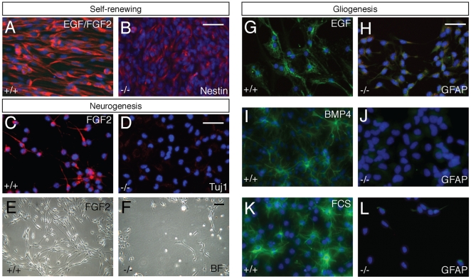
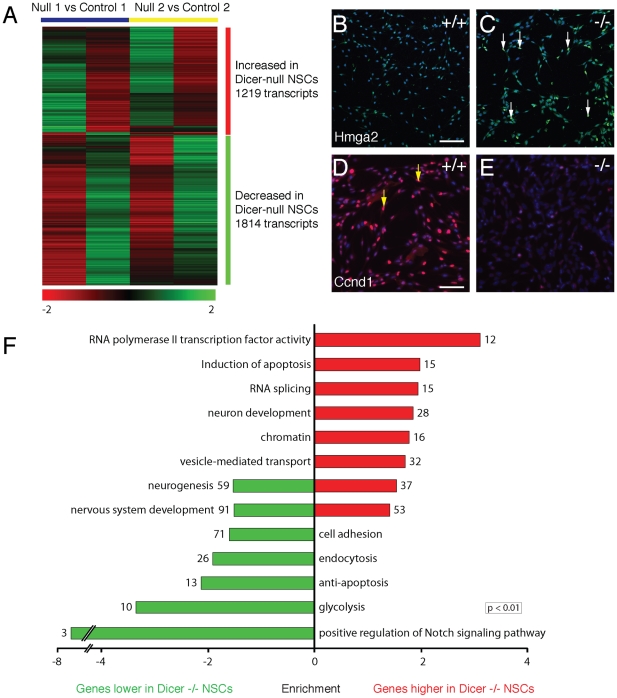
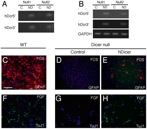
Principal findings: Dicer-null NS cells underwent normal self-renewal and could be maintained in vitro indefinitely, but had subtly altered cell cycle kinetics and abnormal heterochromatin organisation. In the absence of all microRNAs, Dicer-null NS cells were incapable of generating either glial or neuronal progeny and exhibited a marked dependency on exogenous EGF for survival. Dicer-null NS cells assumed complex differences in mRNA and protein expression under self-renewing conditions, upregulating transcripts indicative of self-renewing NS cells and expressing genes characteristic of differentiating neurons and glia. Underlining the growth-factor dependency of Dicer-null NS cells, many regulators of apoptosis were enriched in expression in these cells. Dicer-null NS cells initiate some of the same gene expression changes as wild-type cells under astrocyte differentiating conditions, but also show aberrant expression of large sets of genes and ultimately fail to complete the differentiation programme. Acute replacement of Dicer restored their ability to differentiate to both neurons and glia.

Conclusions: The block in differentiation due to loss of Dicer and microRNAs is reversible and the significantly altered phenotype of Dicer-null NS cells does not constitute a permanent transformation. We conclude that Dicer and microRNAs function in this system to maintain the neural stem cell phenotype and to facilitate the completion of differentiation.


Conflict of interest statement

Competing Interests: The authors have declared that no competing interests exist.

Figures

Figure 1
Figure 1. Dicer-null cortical neural stem cells can be established from the embryonic cerebral cortex and self-renewal appropriately.
(A) At E15, several days after Dicer ablation using the neural progenitor Nes-Cre deleter line, Dicer-null cortices still have about 40% of wild-type microRNA levels (average of the relative expression of all microarray–detected microRNAs), whereas microRNAs are almost undetectable in the cortex at birth. MicroRNA levels were measured as described in the methods and Table S1. (B) PCR genotyping of monolayer cultures of cortex-derived NS cell lines demonstrates that lines derived from Nes-Cre;Dicerfl/fl E12 cortex are Cre+ and contain only the recombined Dicer allele. (C) Dicer-null NS cells lack expression of microRNAs, compared to wild-type cells, although expression of other short RNAs, such as tRNAs, is normal. Expression for a subset of typical microRNAs and two tRNAs is shown relative to expression in control NS cells, and is the average of six replicate features on the array. (D–G) Neural stem cell lines established from wild-type and Dicer-null E12.5 cortex in monolayer culture both express the NS-cell specific intermediate filament protein Nestin (D, E) and the NS cell transcription factor Sox2 (F, G). While Nestin is clearly expressed, the morphology of Dicer-null NS cells is abnormal and lacks the long radial fibres found in wild-type NS cells. Scale bars, 25 µm. (H, I) Dicer-null and control NS cells both form neurospheres in non-adherent culture, in which the majority of cells express Nestin. Note that cells at the periphery of neurospheres generated by control cells begin to differentiate to astrocytes, as indicated by GFAP expression, whereas no GFAP expression is observed in Dicer-null neurospheres. Scale bar, 25 µm. Nuclei in all images visualised with DAPI.
Figure 2
Figure 2. Dicer-null NS cells have minor alterations cell cycle kinetics and significant changes in heterochromatin organisation.
(A–D) Plots of the cell cycle distributions of wildtype (A, B) and Dicer-null (C, D) cells. In both genotypes the majority (∼60%) of cells are in G1, reflecting the relatively slow cycle times for NS cells. Y-axis, cell counts; x-axis, DNA content (Hoechst 33342 fluorescence). (E) Histogram of the proportions of NS cells in each phase of the cell cycle. There is a significant difference in the numbers of cells in G1 in the two control lines, however there is a consistent decrease in the proportion of Dicer-null NS cells in G2/M, compared with controls. (F, G) High-power, confocal images of wild-type (F) and Dicer-null (G) NS cells stained for H3K9Me3. Representative images are shown of the distribution of H3K9Me3 staining in control cells as a relatively small number of large foci (F, arrows). In contrast, the H3K9Me3 distribution is variable in the Dicer-null NS cells, with the majority of cells containing smaller, irregular foci of H3K9Me3 (G). Scale bar, 10 µm. (H) Dicer-null NS cells are karotypically normal, containing the normal complement of 40 chromosomes for mouse and no obvious translocations.
Figure 3
Figure 3. Dicer-null NS cells are incapable of neurogenesis and gliogenesis.
(A, B) Under self-renewing conditions, Dicer-null NS cells express Nestin, but at a lower level than control NS cells. (C–F) Withdrawal of EGF and continued exposure to FGF2 promotes neurogenesis in control NS cells (Tuj1+ cells, C), but does not result in neurogenesis in Dicer-null NS cells (D). Shortly after EGF withdrawal most Dicer-null NS cells die and detach from the culture plate. Brightfield image of wild-type (E) and Dicer-null (F) 24 hours after EGF-withdrawal. (G, H) Withdrawal of FGF2 and continued exposure to EGF promotes gliogenesis in control NS cells (GFAP+ cells, G), but does not result in gliogenesis in Dicer-null NS cells (H), based on both GFAP expression and cell morphology. (I, J) Control NS cells generate large numbers of GFAP+ astrocytes 48 hours after exposure to BMP4 (I), whereas Dicer-null NS cells fail to express GFAP or assume an astrocytic morphology (J). (K, L) Addition of foetal calf serum (FCS) promotes gliogenesis from control cells within 48 hours (K), but does not stimulate gliogenesis from Dicer-null NS cells (L). Scale bar for all images, 25 µm. Nuclei in all images visualised with DAPI.
Figure 4
Figure 4. Dicer-null NS cells exhibit complex changes in mRNA levels resulting in a substantially altered phenotype.
(A) Dicer-null NS cells show significant changes in the mRNA levels of 3033 genes, compared with control NS cells. Although a small number of transcripts demonstrated over ten-fold alterations in expression, the overwhelming majority showed changes of two-fold or less. See methods for technical details of the array hybridisations and statistical analysis. Detailed gene lists are provided in Table S2. (B–E) Changes in mRNA transcripts were reflected in changes in protein expression in cultures of Dicer-null NS cells. Increased Hmga2 mRNA was found to result in the appearance of a subset of strongly HMGA2-expressing nuclei in Dicer-null NS cells, as indicated by the arrows (B, C). Reduced levels of Cyclin D1 mRNA was reflected in the absence of CyclinD1 protein-positive Dicer-null NS cells, compared with controls (yellow arrows in D, with no Ccnd1+ cells detected in the Dicer-null cells in E). Antibody staining is representative of that seen in at least two cultures in two independent lines for each genotype. Scale bar, 25 µm. Nuclei are visualised with DAPI counterstaining. (F) Key Gene Ontology categories found enriched in the sets of genes up (red) and down-regulated in Dicer-null NS cells, compared to wild-type controls. For each category, the number represents the number of genes identified in that category, and the bar size reflects the fold-enrichment of that category in the up- or down-regulated gene set compared with the whole probeset. Details of all enriched GO categories are provided in Table S3.
Figure 5
Figure 5. Dicer-null NS cells initiate but fail to complete a differentiation program.
(A–D) Following two days of astrocytic differentiation in response to BMP4, Nestin protein is down-regulated in both control and Dicer-null NS cells, whereas the expression of the astrocyte-specific protein GFAP is only induced in control NS cells. Scale bar, 25 µm. (E–H) Microarray analysis of BMP4-induced glial differentiation in control and Dicer-null NS cells. Examples of the four major patterns of gene expression observed within 24 hours of BMP4 exposure are shown. In each cluster, each column represents a microarray experiment, each row a unique gene. Two hybridisations for each comparison are shown: control cells 24 hours after BMP4 exposure compared with untreated control cells (+/+); Dicer-null cells 24 hours after BMP4 exposure compared with untreated Dicer-null cells (−/−); BMP4-treated wild-type compared with BMP4-treated Dicer-null cells (+/+ vs −/−). Clusters of transcripts upregulated in both cell types (E) and down-regulated in both cell types (F) are shown, demonstrating that Dicer-null NS cells do execute components of the astrocytic differentiation program. However, as shown in (G) Dicer-null cells also upregulate a set of genes to an inappropriately high level, and also fail to upregulate a large set of genes that are upregulated in control cells (H). (I) Significance analysis of microarrays (SAM) plot comparing gene expression between BMP4-treated wild-type and Dicer-null NS cells (see methods for details). At a 0.1 false discovery rate, two sets of transcripts showing differential expression between the two groups of arrays (BMP4-treated Dicer-null/untreated Dicer-null vs BMP4-treated wild-type/untreated wild-type) were observed: genes more highly expressed in BMP-treated Dicer-null cells compared with the equivalent wild-type cells, and genes more highly expressed in BMP4-treated wild-type cells compared with the equivalent Dicer-null cells. (J, K) Hierarchical clustering of the two sets of transcripts detected by the SAM analysis reported in (I). Within each set, two subclusters were found and are labeled: transcripts higher in BMP4-treated Dicer-null cells contained a cluster of genes down-regulated only in the wild-type cells, but not in the Dicer-nulls (J), and a cluster of genes up-regulated in the Dicer-null cells, but not in the wild-type cells (J); similarly, transcripts higher in BMP4-treated wild-type cells contained a cluster of genes up-regulated only in the wild-type cells, but not in the Dicer-nulls (K), and a small cluster of genes down-regulated in the Dicer-null cells, but not in the wild-type cells (K). Cluster contents are detailed in Table S4.
Figure 6
Figure 6. The differentiation block in Dicer-null NS cells can be reversed by reintroduction of Dicer.
(A) Genomic PCR for human Dicer (hDicer) with two different sets of hDicer-specific primers in two lines of Dicer-null NS cells following infection with a luciferase-expressing virus (control, C) or a hDicer-expressing virus (hD) demonstrated the presence of hDicer DNA in the pFB-hDicer infected cells. (B) RT-PCR for hDicer RNA in virally-infected cells demonstrates expression of hDicer mRNA. (C–E) Wildtype control cells (C) differentiate to GFAP-expressing astrocytes 48 hours after exposure to FCS, in contrast with Dicer-null NS cells infected with a luciferase-expressing virus (D). Dicer-null NS cells infected with a hDicer-containing virus 24 hours before FCS addition recover the ability to generate GFAP-expressing astrocytes (E). (F–H) Similarly, wildtype cells (F) differentiate to Tuj1-expressing neurons upon withdrawal of EGF and maintenance of FGF2, in contrast with Dicer-null NS cells infected with a luciferase-expressing virus (G), the majority of which undergo apoptosis. Dicer-null NS cells infected with the hDicer-containing virus recover the ability to generate Tuj1-expressing neurons (H). Scale bar, 25 µm. Nuclei in all panels visualised with DAPI.

Similar articles

Cited by

References

    1. Bartel DP, Chen CZ. Micromanagers of gene expression: the potentially widespread influence of metazoan microRNAs. Nat Rev Genet. 2004;5:396–400. - PubMed
    1. Ambros V. The functions of animal microRNAs. Nature. 2004;431:350–355. - PubMed
    1. Kosik KS, Krichevsky AM. The Elegance of the MicroRNAs: A Neuronal Perspective. Neuron. 2005;47:779–782. - PubMed
    1. Cheng LC, Tavazoie M, Doetsch F. Stem cells: from epigenetics to microRNAs. Neuron. 2005;46:363–367. - PubMed
    1. Lagos-Quintana M, Rauhut R, Yalcin A, Meyer J, Lendeckel W, et al. Identification of tissue-specific microRNAs from mouse. Curr Biol. 2002;12:735–739. - PubMed

Publication types