

Betacellulin induces increased retinal vascular permeability in mice
- PMID: 20976146
- PMCID: PMC2956654
- DOI: 10.1371/journal.pone.0013444
Betacellulin induces increased retinal vascular permeability in mice
Abstract
Background: Diabetic maculopathy, the leading cause of vision loss in patients with type 2 diabetes, is characterized by hyper-permeability of retinal blood vessels with subsequent formation of macular edema and hard exudates. The degree of hyperglycemia and duration of diabetes have been suggested to be good predictors of retinal complications. Intervention studies have determined that while intensive treatment of diabetes reduced the development of proliferative diabetic retinopathy it was associated with a two to three-fold increased risk of severe hypoglycemia. Thus we hypothesized the need to identify downstream glycemic targets, which induce retinal vascular permeability that could be targeted therapeutically without the additional risks associated with intensive treatment of the hyperglycemia. Betacellulin is a 32 kD member of the epidermal growth factor family with mitogenic properties for the retinal pigment epithelial cells. This led us to hypothesize a role for betacellulin in the retinal vascular complications associated with diabetes.
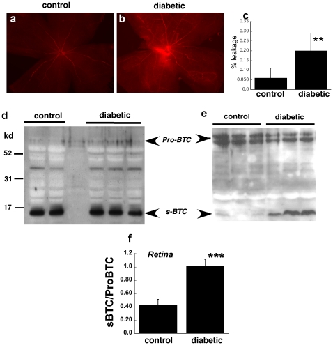
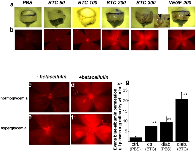
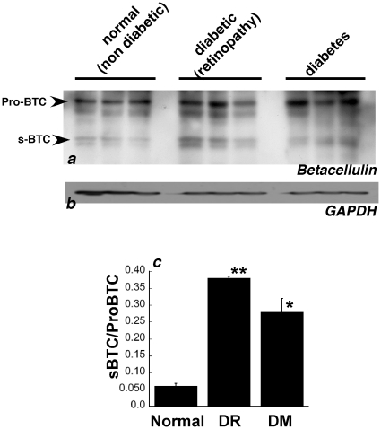
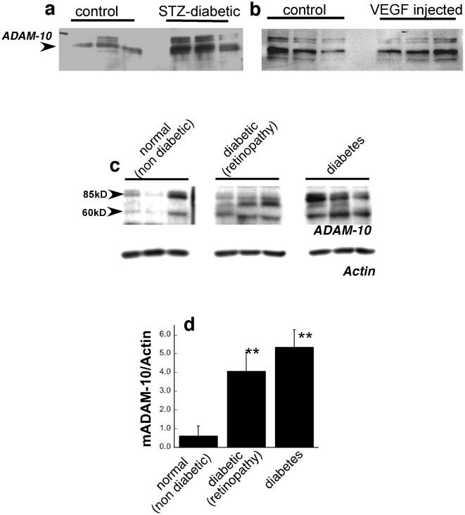
Methods and findings: In this study, using a mouse model of diabetes, we demonstrate that diabetic mice have accentuated retinal vascular permeability with a concomitant increased expression of a cleaved soluble form of betacellulin (s-Btc) in the retina. Intravitreal injection of soluble betacellulin induced retinal vascular permeability in normoglycemic and hyperglycemic mice. Western blot analysis of retinas from patients with diabetic retinopathy showed an increase in the active soluble form of betacellulin. In addition, an increase in the levels of A disintegrin and metalloproteinase (ADAM)-10 which plays a role in the cleavage of betacellulin was seen in the retinas of diabetic mice and humans.
Conclusions: These results suggest that excessive amounts of betacellulin in the retina may contribute to the pathogenesis of diabetic macular edema.
Conflict of interest statement
Figures
Similar articles
-
Inhibition of EGF signaling protects the diabetic retina from insulin-induced vascular leakage.Am J Pathol. 2013 Sep;183(3):987-95. doi: 10.1016/j.ajpath.2013.05.017. Epub 2013 Jul 3. Am J Pathol. 2013. PMID: 23831329 Free PMC article.
-
Decreased lysyl oxidase level protects against development of retinal vascular lesions in diabetic retinopathy.Exp Eye Res. 2019 Jul;184:221-226. doi: 10.1016/j.exer.2019.04.019. Epub 2019 May 6. Exp Eye Res. 2019. PMID: 31022398 Free PMC article.
-
Vascular permeability in retinopathy is regulated by VEGFR2 Y949 signaling to VE-cadherin.Elife. 2020 Apr 21;9:e54056. doi: 10.7554/eLife.54056. Elife. 2020. PMID: 32312382 Free PMC article.
-
Regulation of retinal vascular permeability by betacellulin.Adv Exp Med Biol. 2012;723:293-8. doi: 10.1007/978-1-4614-0631-0_38. Adv Exp Med Biol. 2012. PMID: 22183345 Review. No abstract available.
-
[Cell biology of intraocular vascular diseases].Nippon Ganka Gakkai Zasshi. 1999 Dec;103(12):923-47. Nippon Ganka Gakkai Zasshi. 1999. PMID: 10643294 Review. Japanese.
Cited by
-
Effect of Lentivirus-Mediated miR-499a-3p on Human Umbilical Vein Endothelial Cells.Biomed Res Int. 2020 Aug 26;2020:9372961. doi: 10.1155/2020/9372961. eCollection 2020. Biomed Res Int. 2020. PMID: 32908925 Free PMC article.
-
Preliminary identification of pathogenic variants in an Afro-Colombian Raizal family with risk factors for glaucoma.Colomb Med (Cali). 2022 Jun 30;53(2):e2065107. doi: 10.25100/cm.v53i2.5107. eCollection 2022 Apr-Jun. Colomb Med (Cali). 2022. PMID: 36415692 Free PMC article.
-
SOCS1-Derived Peptide Administered by Eye Drops Prevents Retinal Neuroinflammation and Vascular Leakage in Experimental Diabetes.Int J Mol Sci. 2019 Jul 24;20(15):3615. doi: 10.3390/ijms20153615. Int J Mol Sci. 2019. PMID: 31344857 Free PMC article.
-
Gene expression changes during retinal development and rod specification.Mol Vis. 2015 Jan 20;21:61-87. eCollection 2015. Mol Vis. 2015. PMID: 25678762 Free PMC article.
-
Mechanistic Insights into Pathological Changes in the Diabetic Retina: Implications for Targeting Diabetic Retinopathy.Am J Pathol. 2017 Jan;187(1):9-19. doi: 10.1016/j.ajpath.2016.08.022. Epub 2016 Nov 12. Am J Pathol. 2017. PMID: 27846381 Free PMC article. Review.
References
-
- Group TDCacTR. Implications of the Diabetes Control and Complications Trial. American Diabetes Association. Diabetes. 1993;42:1555–1558. - PubMed
-
- Keenan HA, Costacou T, Sun JK, Doria A, Cavellerano J, et al. Clinical factors associated with resistance to microvascular complications in diabetic patients of extreme disease duration: the 50-year medalist study. Diabetes Care. 2007;30:1995–1997. - PubMed
-
- Group. TDCaCTR. The effect of intensive treatment of diabetes on the development and progression of long-term complications in insulin-dependent diabetes mellitus. The Diabetes Control and Complications Trial Research Group. N Engl J Med. 1993;329:977–986. - PubMed
Publication types
MeSH terms
Substances
Grants and funding
LinkOut - more resources
Full Text Sources
Other Literature Sources
