. 2010 Oct 14;6(10):e1001144.
doi: 10.1371/journal.ppat.1001144.

Early production of IL-22 but not IL-17 by peripheral blood mononuclear cells exposed to live Borrelia burgdorferi: the role of monocytes and interleukin-1

Affiliations

Early production of IL-22 but not IL-17 by peripheral blood mononuclear cells exposed to live Borrelia burgdorferi: the role of monocytes and interleukin-1

Malte Bachmann et al. PLoS Pathog. .

Abstract

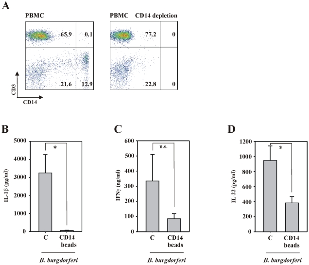
If insufficiently treated, Lyme borreliosis can evolve into an inflammatory disorder affecting skin, joints, and the CNS. Early innate immunity may determine host responses targeting infection. Thus, we sought to characterize the immediate cytokine storm associated with exposure of PBMC to moderate levels of live Borrelia burgdorferi. Since Th17 cytokines are connected to host defense against extracellular bacteria, we focused on interleukin (IL)-17 and IL-22. Here, we report that, despite induction of inflammatory cytokines including IL-23, IL-17 remained barely detectable in response to B. burgdorferi. In contrast, T cell-dependent expression of IL-22 became evident within 10 h of exposure to the spirochetes. This dichotomy was unrelated to interferon-γ but to a large part dependent on caspase-1 and IL-1 bioactivity derived from monocytes. In fact, IL-1β as a single stimulus induced IL-22 but not IL-17. Neutrophils display antibacterial activity against B. burgdorferi, particularly when opsonized by antibodies. Since neutrophilic inflammation, indicative of IL-17 bioactivity, is scarcely observed in Erythema migrans, a manifestation of skin inflammation after infection, protective and antibacterial properties of IL-22 may close this gap and serve essential functions in the initial phase of spirochete infection.


Conflict of interest statement

The authors have declared that no competing interests exist.

Figures

Figure 1
Figure 1. A specific cytokine pattern associated with PBMC exposed to live B. burgdorferi.
(A) PBMC were either kept as unstimulated control or exposed to B. burgdorferi 297 (MOI = 0.1). After 65 h cell-free cell culture supernatants were analyzed in a 1∶3 dilution by antibody array analysis. One representative of two independently performed experiments (two different donors) is shown. (B) PBMC were either kept as unstimulated control or exposed to the indicated MOI of B. burgdorferi 297. After 65 h, TNFα release was determined by ELISA. Data are expressed as means ± SEM (n = 6 for unstimulated control and MOI = 0.1, n = 3 for MOI = 1 and 3); **p<0.01, ***p<0.001 compared with unstimulated control; raw data were analyzed by one-way ANOVA with post hoc Bonferroni correction. (C) PBMC were either kept as unstimulated control or were exposed to B. burgdorferi 297 (MOI = 0.1). After the indicated time periods TNFα release was determined by ELISA. Data are expressed as means ± SEM (n = 3); **p<0.01, compared with unstimulated control at the respective time point; raw data were analyzed by unpaired Student's t-test. (D-I) PBMC were either kept as unstimulated control or were exposed to B. burgdorferi 297 (MOI = 0.1). After 24 h (D-G, I) or 65 h (H), release of IL-1β (D; n = 3; *p<0.05, compared with unstimulated control), IL-23 (E; n = 3; *p<0.05, compared with unstimulated control), IL-12 (F; n = 5; ***p<0.001, compared with unstimulated control), IFNγ (G; n = 3; ***p<0.001, compared with unstimulated control), IL-8 (H; n = 5; ***p<0.001, compared with unstimulated control; where indicated experiments were performed in the presence of PmxB at 3 µg/ml), and IL-18 (I; n = 3; *p<0.05, compared with unstimulated control) was determined by ELISA. Raw data were either analyzed by unpaired Student's t-test (D-G, I) or by one-way ANOVA with post hoc Bonferroni correction (H).
Figure 2
Figure 2. Production of IL-22 but not of IL-17 by PBMC exposed to live B. burgdorferi.
(A) PBMC were either kept as unstimulated control or exposed to the indicated MOI of B. burgdorferi 297. After 65 h, IL-22 (closed triangles), IL-17 (closed circles), and IL-17F (open circles) release was determined by ELISA. Data are expressed as means ± SEM (n = 10); **p<0.01, compared with unstimulated control; raw data were analyzed by one-way ANOVA with post hoc Bonferroni correction. (B) PBMC were either kept as unstimulated control or were exposed to B. burgdorferi 297 (MOI = 0.1). After the indicated incubation periods, total RNA was isolated and IL-22 mRNA expression was determined by real-time PCR analysis. IL-22 mRNA was normalized to that of GAPDH and is shown as mean fold induction compared to unstimulated control (at the respective time point) ± SEM (n = 3); *p<0.05, **p<0.01, ***p<0.001 compared with unstimulated control (at the respective time point); raw data were analyzed by unpaired Student's t-test. Inset: In addition to real-time PCR, data from one representative experiment was analyzed by standard PCR analysis. (C) PBMC were either kept as unstimulated control or were exposed to B. burgdorferi 297 (MOI = 0.1). After the indicated time periods IL-22 release was determined by ELISA. Data are expressed as means ± SEM (n = 5); **p<0.01, ***p<0.001 compared with unstimulated control at the respective time point; raw data were analyzed by unpaired Student's t-test. (D) Jurkat T cells were transfected for 5 h with pGL3-IL22 together with Renilla luciferase as described in the materials and methods section. After 15 h of rest, cells were resuspended in control conditioned media (CCM) obtained from unstimulated PBMC or in conditioned media from PBMC exposed to B. burgdorferi 297 (either MOI = 0.1 (n = 3) or MOI = 10 (n = 6), stimulation period: 15 h) and stimulated with αCD3 (25 µg/ml) thereafter. After another 8 h, cells were harvested and luciferase assays were performed. Data are expressed as promoter activity (% of αCD3 at CCM ± SD); **p<0.01, ***p<0.001 compared with αCD3 at CCM; #p<0.05, ###p<0.001 compared with Jurkat T cells in CCM in the absence of αCD3 and B. burgdorferi. Raw data were analyzed by one-way ANOVA with post hoc Bonferroni correction. (E) Jurkat T cells were treated using the protocol described under (D). However, only B. burgdorferi at a MOI of 10 were used to generate PBMC-derived conditioned media. In addition, αCD3 stimulation of Jurkat T cells was omitted. Data are expressed as promoter activity (% of CCM ± SD) (n = 8); ***p<0.001 compared with promoter activity at CCM. Raw data were analyzed by unpaired Student's t-test. (FG) PBMC were exposed to B. burgdorferi 297 (MOI = 0.1) either alone or in combination with the indicated concentrations of IL-10 (F) or dexamethasone (G). After 65 h, IL-22 release was determined by ELISA. Data (% of B. burgdorferi alone) are expressed as means ± SEM (IL-10: n = 6, dexamethasone: n = 3); closed circles denote unstimulated control, closed squares denote IL-10 at 20 ng/ml (F) or dexamethasone at 50 nM (G) alone; **p<0.01 compared with unstimulated control; #p<0.05, ##p<0.01 compared with PBMC exposed to B. burgdorferi 297 alone; raw data were analyzed by one-way ANOVA with post hoc Bonferroni correction. (H) PBMC were either kept as unstimulated control or stimulated with IL-2 (20 ng/ml)/IL-23 (20 ng/ml) or were exposed to B. burgdorferi 297 (MOI = 0.1). After 65 h, IL-17 release was determined by ELISA. Data are expressed as means ± SEM (n = 15 for control and IL-2/IL-23, n = 8 for B. burgdorferi 297); n.s. denotes not significantly different from unstimulated control; ***p<0.001, compared with unstimulated control, ##p<0.01 compared with IL-2/IL-23; raw data were analyzed by one-way ANOVA with post hoc Bonferroni correction. (I) PBMC were either kept as unstimulated control or were exposed to the indicated dosages of B. burgdorferi 297. After 42 h, IL-10 release was determined by ELISA. Data are expressed as means ± SEM (n = 8 for control and B. burgdorferi 297 at a MOI of 0.1, n = 6 for B. burgdorferi 297 at a MOI of 10); n.s. denotes not significantly different from unstimulated control; **p<0.01, compared with unstimulated control; raw data were analyzed by one-way ANOVA with post hoc Bonferroni correction.
Figure 3
Figure 3. IL-1 drives production of IL-22 by PBMC exposed to live B. burgdorferi.
(A) PBMC were kept as unstimulated control or cultivated together with IL-1Ra (1.3 µg/ml) in the presence or absence of B. burgdorferi 297 (MOI = 0.1). After 65 h, IL-22 release was determined by ELISA. Data are expressed as means ± SEM (n = 5); **p<0.01 compared with unstimulated control, #p<0.05 compared with B. burgdorferi 297/IL-1Ra. (B) PBMC were kept as unstimulated control or cultivated together with Ac-YVAD-CHO (50 µM) in the presence or absence of B. burgdorferi 297 (MOI = 0.1). After 48 h, IL-22 release was determined by ELISA. Data are expressed as means ± SEM (n = 12); ***p<0.001 compared with unstimulated control, #p<0.05 compared with B. burgdorferi 297/Ac-YVAD-CHO. (AB) IL-1Ra and Ac-YVAD-CHO were added to the cultures 0.5 h before exposure of PBMC to B. burgdorferi. (C) PBMC were either kept as unstimulated control or stimulated with IL-1β (50 ng/ml). After 65 h, IL-22 (black bars) and IL-17 (grey bars) release was determined by ELISA. Data are expressed as means ± SEM (n = 7); **p<0.01, compared with unstimulated control, ##p<0.01 compared with IL-17 levels in the presence of IL-1β. (ABC) Raw data were analyzed by one-way ANOVA with post hoc Bonferroni correction.
Figure 4
Figure 4. T cell-dependent production of IL-22 by PBMC exposed to live B. burgdorferi.
After isolation, PBMC were either depleted for CD3+ (A) or CD56+ (B) cells (see methods section). FACS analysis was performed by gating on lymphocytes in order to confirm successful manipulation of PBMC. Representative depletion experiments are shown. Thereafter, whole PBMC and either CD3 (C) or CD56 (D) depleted cell populations were exposed to B. burgdorferi 297 (MOI = 0.1). After 65 h, release of IL-22 and IFNγ was determined by ELISA. Data are expressed as means ± SEM (CD3 depletion: n = 5; CD56 depletion: n = 8); n.s. denotes not significantly different from B. burgdorferi 297-stimulated whole PBMC; *p<0.05 compared with B. burgdorferi 297-stimulated whole PBMC. Raw data were analyzed by unpaired Student's t-test.
Figure 5
Figure 5. IL-22 expression by PBMC exposed to live B. burgdorferi is dependent on both, CD4+ and CD8+ T cells.
After isolation, PBMC were either depleted for CD4+ (A) or CD8+ (B) cells (see methodssection). FACS analysis was performed by gating on lymphocytes in order to confirm successful manipulation of PBMC. Representative depletion experiments are shown. (C) Thereafter, whole PBMC and either CD4 or CD8 depleted cell populations were kept as unstimulated control or exposed to B. burgdorferi 297 (MOI = 0.1). After 65 h, release of IL-22 was determined by ELISA. Data are expressed as means ± SEM (n = 3); ***p<0.001 compared with unstimulated whole PBMC; ## p< 0.01, ### p<0.001 compared with whole PBMC exposed to B. burgdorferi 297. Raw data were analyzed by one-way ANOVA with post hoc Bonferroni correction. (D) PBMC from three different donors were either kept as unstimulated control or were exposed to B. burgdorferi 297 (MOI = 0.1). After 24 h, CD4+ and CD8+ T cells were isolated as outlined in the materials and methods section. After isolation of total RNA from either unfractionated PBMC and CD4+ or CD8+ T cells, IL-22 mRNA expression was analyzed by standard PCR. Data from all three donors are shown.
Figure 6
Figure 6. Production of IL-22 by PBMC exposed to live B. burgdorferi partially depends on intermediate monocyte activation.
(A) After isolation, PBMC were depleted for CD14+ cells (see materials and methods section). FACS analysis was performed in order to confirm successful manipulation of PBMC. Representative depletion experiments are shown. Thereafter, whole PBMC or CD14-depleted cell populations were exposed to B. burgdorferi 297 (MOI = 0.1). After 65 h, release of IL-1β (B), IFNγ (C), and IL-22 (D) was determined by ELISA. Data are expressed as means ± SEM (n = 5); n.s. denotes not significantly different from B. burgdorferi 297-stimulated whole PBMC; *p<0.05 compared with B. burgdorferi 297-stimulated whole PBMC. Raw data were analyzed by unpaired Student's t-test.
Figure 7
Figure 7. Activation of pDC by CpG oligonucleotides does not associate with enhanced production of IL-22 by PBMC.
PBMC were either kept as unstimulated control, were exposed to B. burgdorferi 297 (MOI = 0.1) or were activated by CpG oligonucleotides (10 µg/ml) type A (A) or type C (B) alone or in the presence of B. burgdorferi 297 (MOI = 0.1). After 65 h, release of IL-22 (left panel) and IFNα (right panel) was determined by ELISA. Data are expressed as means ± SEM (n = 6); *p<0.05, ***p<0.001 compared with unstimulated control; ## p<0.01, ### p<0.001 compared with B. burgdorferi 297 alone. Raw data were analyzed by one-way ANOVA with post hoc Bonferroni correction.
Figure 8
Figure 8. Analysis of IL-22 expression in biopsy specimens obtained from erythema chronicum migrans patients.
7 out of 12 specimens obtained from erythema chronicum migrans patients displayed abundant presence of IL-22 positive cells. Two of these skin biopsies are shown in two magnifications.

Similar articles

Cited by

References

    1. Aujla SJ, Kolls JK. IL-22: a critical mediator in mucosal host defense. J Mol Med. 2009;87:451–454. - PubMed
    1. Wolk K, Witte E, Witte K, Warszawska K, Sabat R. Biology of interleukin-22. Semin Immunopathol. 2010;32:17–31. - PubMed
    1. Gaffen SL. An overview of IL-17 function and signaling. Cytokine. 2008;43:402–407. - PMC - PubMed
    1. Miossec P, Korn T, Kuchroo VK. Interleukin-17 and type 17 helper T cells. N Engl J Med. 2009;361:888–898. - PubMed
    1. Steinman L. A brief history of T(H)17, the first major revision in the T(H)1/T(H)2 hypothesis of T cell-mediated tissue damage. Nat Med. 2007;13:139–145. - PubMed

Publication types

MeSH terms