. 2010 Oct 14;6(10):e1001148.
doi: 10.1371/journal.ppat.1001148.

Leishmania-induced inactivation of the macrophage transcription factor AP-1 is mediated by the parasite metalloprotease GP63

Affiliations

Leishmania-induced inactivation of the macrophage transcription factor AP-1 is mediated by the parasite metalloprotease GP63

Irazú Contreras et al. PLoS Pathog. .

Abstract

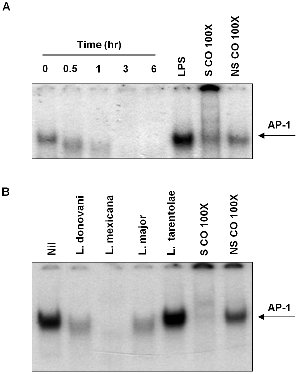
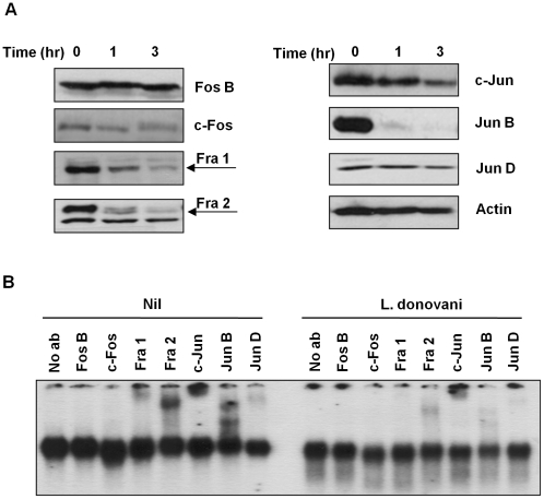
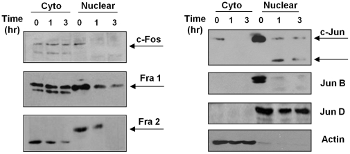
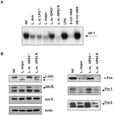
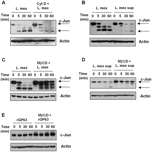
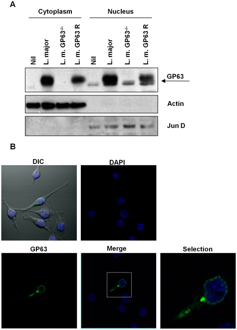
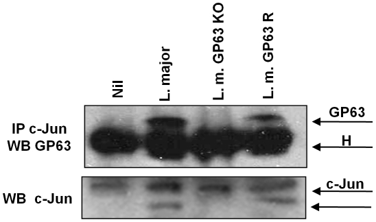
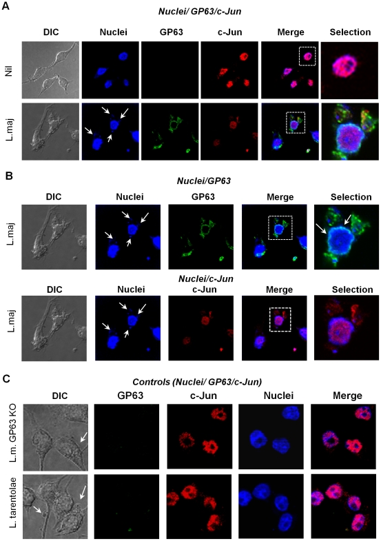
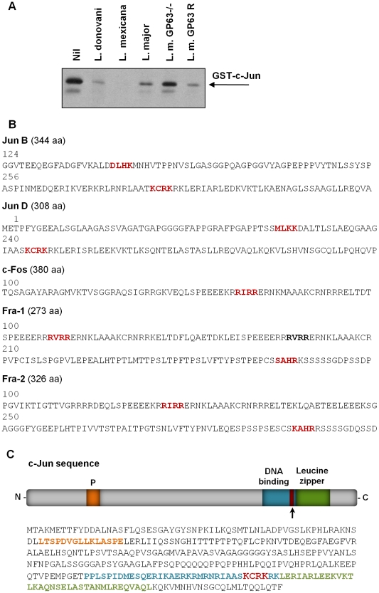
Leishmania parasites have evolved sophisticated mechanisms to subvert macrophage immune responses by altering the host cell signal transduction machinery, including inhibition of JAK/STAT signalling and other transcription factors such as AP-1, CREB and NF-κB. AP-1 regulates pro-inflammatory cytokines, chemokines and nitric oxide production. Herein we show that upon Leishmania infection, AP-1 activity within host cells is abolished and correlates with lower expression of 5 of the 7 AP-1 subunits. Of interest, c-Jun, the central component of AP-1, is cleaved by Leishmania. Furthermore, the cleavage of c-Jun is dependent on the expression and activity of the major Leishmania surface protease GP63. Immunoprecipitation of c-Jun from nuclear extracts showed that GP63 interacts, and cleaves c-Jun at the perinuclear area shortly after infection. Phagocytosis inhibition by cytochalasin D did not block c-Jun down-regulation, suggesting that internalization of the parasite might not be necessary to deliver GP63 molecules inside the host cell. This observation was corroborated by the maintenance of c-Jun cleavage upon incubation with L. mexicana culture supernatant, suggesting that secreted, soluble GP63 could use a phagocytosis-independent mechanism to enter the host cell. In support of this, disruption of macrophage lipid raft microdomains by Methyl β-Cyclodextrin (MβCD) partially inhibits the degradation of full length c-Jun. Together our results indicate a novel role of the surface protease GP63 in the Leishmania-mediated subversion of host AP-1 activity.


Conflict of interest statement

The authors have declared that no competing interests exist.

Figures

Figure 1
Figure 1. Infection with different species of Leishmania inhibits AP-1 DNA binding activity.
(A) B10R macrophages were infected for 0.5, 1, 3 and 6 hr with L. donovani, at a ratio 20∶1 parasite/macrophage. Nuclear proteins were isolated and EMSA for AP-1 DNA binding activity was performed. A consensus DNA sequence for SP-1 binding was used as non-specific competitor (NSCO). A 100× molar excess of AP-1 probe was used as a specific competitor (SCO). (B) B10R macrophages were infected for 1 hr with L. mexicana, L. major, L donovani infantum or L. tarentolae and treated as in (A).
Figure 2
Figure 2. AP-1 subunits are degraded after infection with Leishmania parasites.
(A) Western blot analysis of AP-1 subunit proteins extracted from B10R macrophages infected with L. donovani for 1 and 3 hr. β-actin was used as a loading control. (B) Super shift assays of B10R macrophages infected with L. donovani for 1 hr. Nuclear proteins were super shifted using antibodies against c-Jun, Jun B, Jun D, c-Fos, Fos B, Fra 1 and Fra 2 AP-1 subunits.
Figure 3
Figure 3. Subcellular localization of AP-1 subunits.
B10R macrophages were infected with L. donovani for 1 and 3 hr. Cytoplasmic and nuclear distribution of the AP-1 subunits was monitored by Western Blot analysis. β-actin was used as a loading control for cytoplasmic fraction, and Jun D for nuclear fraction.
Figure 4
Figure 4. Role of Leishmania surface molecules in the inactivation of AP-1.
(A) EMSA for AP-1 DNA binding activity of nuclear extracts from B10R macrophages infected for 1 hr with L. donovani 1S2D (LPG+/+), L. donovani LPG−/−, L. major (WT), L. major GP63 −/− or L. major GP63 Rescued (GP63 R) promastigotes. A consensus DNA sequence for SP-1 binding was used as non-specific control (NSCO). A 100× molar excess of AP-1 probe was used as a specific competitor (SCO). 1 hr stimulation with LPS (100 ng/ml) was used as a positive control for the induction of AP-1 DNA binding. (B) Macrophages were infected for 1 hr with L. major (WT), L. major GP63 −/− or L. major Rescued promastigotes at 20∶1 ratio. WB of AP-1 subunits was performed with the total cell lysate. β-actin was used as a loading control.
Figure 5
Figure 5. GP63 delivery into the host cell is mediated via lipid raft.
(A) B10R macrophages were pre-treated or not with 2 µM cytochalasin D for 1 hr and then infected with L. mexicana for indicated times. (B) B10R macrophages were incubated with either with whole parasite or culture supernatant of L. mexicana promastigotes. Macrophages were pre-treated or not with 20 mM of methyl β-ciclodextrin (MβCD) for 1 hr and infected with L. mexicana promastigotes (C), incubated with L. mexicana supernatant (D) or recombinant GP63 (rGP63) (E). For all the Figures, total cell extracts and β-actin as a loading control were used.
Figure 6
Figure 6. Subcellular localization of GP63.
(A) B10R macrophages were infected for 1 hr with L. major (WT), L. major GP63−/− or L. major GP63 Rescued and GP63 distribution in the cytoplasmic and nuclear extracts was monitored by WB. β-actin and Jun B were used as fractioning controls. (B) B10R macrophages were infected for 1 hr with L. major (WT). GP63 is shown in green and nuclei were stained with DAPI (blue).
Figure 7
Figure 7. c-Jun interacts with GP63 in nuclear fraction.
Proteins from nuclear extracts of macrophages infected for 1 hr with L. major (WT), L. major GP63 −/− and L. major GP63 Rescued (GP63 R) and nuclear proteins were immunoprecipitated using an anti-c-Jun antibody. GP63 and c-Jun co-immunoprecipitation was evaluated by western blot. H denotes the heavy chain of the immunoglobulin.
Figure 8
Figure 8. GP63 is localized in the perinuclear compartment.
(A) Confocal microscopy images of B10R macrophages showing co-localization of nuclei (blue), c-Jun (red) and GP63 (green) in non-infected (upper panel) and cells infected for 1 hr with L. major (lower panel). (B) Confocal microscopy analysis to evaluate nuclear localization of GP63 (green) and nuclear distribution (upper panel) and degradation of c-Jun (red) in macrophages infected 1 hr with L. major (lower panel). Blue shows cell nuclei. (C) co-localization of nuclei (blue), c-Jun (red) and GP63 (green) in B10R macrophages infected for 1 hr with L. major GP63−/− or L. tarentolae.
Figure 9
Figure 9. Parasite-free GP63 is sufficient to degrade c-Jun.
(A) Exogenous GST-c-Jun was incubated with 500 µl of L. donovani, L. mexicana, L. major, L. major GP63−/− or L. major GP63 Rescued for 30 minutes, and degradation of c-Jun was visualized by WB using anti-GST antibody. (B) Jun B, Jun D, c-Fos, Fos B, Fra 1 and Fra 2 protein sequences showing putative GP63 cleavage sites. (C) c-Jun sequence analysis showing the putative site of GP63 cleavage.

Similar articles

Cited by

References

    1. Desjeux P. Leishmaniasis: current situation and new perspectives. Comp Immunol Microbiol Infect Dis. 2004;27:305–318. - PubMed
    1. Piscopo TV, Mallia AC. Leishmaniasis. Postgrad Med J. 2006;82:649–657. - PMC - PubMed
    1. Olivier M, Gregory DJ, Forget G. Subversion mechanisms by which Leishmania parasites can escape the host immune response: a signaling point of view. Clin Microbiol Rev. 2005;18:293–305. - PMC - PubMed
    1. Forget G, Gregory DJ, Olivier M. Proteasome-mediated degradation of STAT1α following infection of macrophages with Leishmania donovani. J Biol Chem. 2005;280:30542–30549. - PubMed
    1. Gregory DJ, Contreras I, Forget G, Olivier M. A novel form of NF-κB is induced by Leishmania infection: Involvement in macrophage gene expression. Eur J Immunol. 2008;38:1071–1081. - PubMed

Publication types

MeSH terms