. 2010 Oct 14;6(10):e1001151.
doi: 10.1371/journal.ppat.1001151.

Epstein Barr virus-encoded EBNA1 interference with MHC class I antigen presentation reveals a close correlation between mRNA translation initiation and antigen presentation

Affiliations

Epstein Barr virus-encoded EBNA1 interference with MHC class I antigen presentation reveals a close correlation between mRNA translation initiation and antigen presentation

Sebastien Apcher et al. PLoS Pathog. .

Abstract

Viruses are known to employ different strategies to manipulate the major histocompatibility (MHC) class I antigen presentation pathway to avoid recognition of the infected host cell by the immune system. However, viral control of antigen presentation via the processes that supply and select antigenic peptide precursors is yet relatively unknown. The Epstein-Barr virus (EBV)-encoded EBNA1 is expressed in all EBV-infected cells, but the immune system fails to detect and destroy EBV-carrying host cells. This immune evasion has been attributed to the capacity of a Gly-Ala repeat (GAr) within EBNA1 to inhibit MHC class I restricted antigen presentation. Here we demonstrate that suppression of mRNA translation initiation by the GAr in cis is sufficient and necessary to prevent presentation of antigenic peptides from mRNAs to which it is fused. Furthermore, we demonstrate a direct correlation between the rate of translation initiation and MHC class I antigen presentation from a certain mRNA. These results support the idea that mRNAs, and not the encoded full length proteins, are used for MHC class I restricted immune surveillance. This offers an additional view on the role of virus-mediated control of mRNA translation initiation and of the mechanisms that control MHC class I restricted antigen presentation in general.


Conflict of interest statement

The authors have declared that no competing interests exist.

Figures
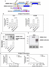
Figure 1
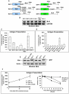
Figure 1. Inhibition of EBNA1 synthesis prevents presentation of peptides derived from the EBNA1 mRNA.
A) Cartoon illustrating the different constructs. The location of the exogenous antigenic peptide sequence SIINFEKL (SL8) of the chicken ovalbumin (Ova) in the EBNA1-SL8 and the EBNA1ΔGA-SL8 constructs is indicated. B) The presentation of SL8 peptide on endogenous MHC class I Kb molecules on (0.5×10⁵) EL4 cells (left) or on human cells co-expressing a genomic Kb construct (right) was determined using B3Z CD8+ T cells . The GAr domain suppresses presentation of SL8 by over 90% in either cell type. C) Autoradiograph of a 1 hour 35S-methionine pulse label in the presence of proteasome inhibitors shows that the EBNA1-SL8 mRNA is translated 60% less efficiently as compared with the EBNA1ΔGA-SL8. The graph below shows values determined from phosphoimager analysis. D) Western blot shows the steady state level of expression of indicated constructs without proteasome inhibitors. E) Dose-response curve shows that approximately 8 µg of GAr-Ova cDNA is required to reach a similar level of SL8 presentation as that of 1 µg of Ova (left panel). Increasing number of EL4 cells expressing indicated constructs in the presence of a fixed amount (5×10⁴) of B3Z (right graph). The results show representative data from at least three independent experiments in which transfected cells were split and tested for protein synthesis or antigen presentation with SD.
Figure 2
Figure 2. Inhibition of protein degradation is not essential for the GAr sequence to prevent endogenous antigen presentation for the MHC class I restricted pathway.
A) The SL8 epitope was inserted in the 3′UTR of the GAr open reading frame (ORF) (GAr-1), in another reading frame (GAr-2) or fused to GAr (GAr-3). The corresponding constructs were also made in which the SL8 was inserted in the GFP mRNA in an identical way (GFP-1 to 3). The AUG, GCC or GGC initiation codons for SL8 in GAr-2 were used. B) Northern blot analysis using the SL8 sequence as probe shows that the GAr sequence does not influence the expression levels of the corresponding mRNAs. C) The GAr suppresses presentation of SL8 throughout the entire mRNA, demonstrating that its capacity to prevent antigen presentation does not depend on controlling protein degradation. D) Changing the initiation codon of the SL8 from AUG to GGC (Glycine) or GCC (Alanine) in the GAr-2 prevents expression and antigen presentation, demonstrating that the SL8 is derived from an individual translation initiation event. Presentation of SL8 expressed as a minigene using the initiation codons AUG, GGC and GCC (SL8AUG, SL8GGC and SL8GCC, respectively). E) Western blot shows the steady state level of expression of indicated constructs (GAr-1 to 3 and GFP-1 to 3). F) IFNγ stimulates antigen presentation by optimising processing of peptides longer than 8–10 residues and has only an effect on antigen presentation when SL8 is expressed in the main ORF and not when expressed as cryptic peptide in an alternative reading frame (left graph). Epoxomicin is a specific proteasome inhibitor and prevents presentation of SL8 when expressed in the main ORF (right graph). This shows that expression of the SL8 epitope as a cryptic peptide does not carry additional residues from the main reading frame. Data are representative of three or more independent experiments and values are shown with SD. Protein synthesis and antigen presentation data are derived from cells transfected with the indicated constructs before split and tested separately.
Figure 3
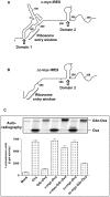
Figure 3. GAr suppresses antigen presentation by targeting the mRNA translation initiation process.
A) Cartoon illustrating the c-myc Internal Ribosomal Entry Sites (IRES) constructs. IRESs offer alternative cap-independent mechanisms of mRNA translation initiation. B) Autoradiograph of 35S-methionine metabolic pulse labelling. Insertion of the c-myc IRES in the 5′UTR of the GAr-Ova mRNA (c-myc-GAr-Ova) restores translation in H1299 cells but does not affect translation when inserted in the 5′UTR of Ova alone (left panel). Western blot shows that the effect of the c-myc IRES is restricted to the GAr alone (right panel). C) Autoradiograph of a 30 minutes 35S-metabolic pulse label experiment of the endogenous protein (actin) and the exogenous GFP protein in H1299 cells in the presence of the c-myc IRES and the GAr constructs. D) The c-myc IRES stimulates SL8 presentation in the context of the GAr from the main open reading frame as well as cryptic translated products (see Fig. 2A). Data are representative of three or more independent experiments plus SD.
Figure 4
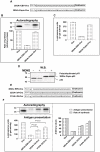
Figure 4. Domain 1 of the c-myc IRES is responsible for the effect of the c-myc IRES on GAr-dependent translation control.
A) Cartoons illustrating the predicted structure and functional domains of the human c-myc IRES and domains 1 and 2 and the ribosome entry window are indicated. B) Domain 1 was deleted while retaining the ribosome entry window and domain 2 (Δc-myc IRES). C) Autoradiograph of 35S-methionine metabolic pulse labelling and presentation of SL8 derived from the indicated constructs in H1299 cells. Insertion of the Δc-myc IRES in the 5′UTR of the GAr-Ova mRNA (Δc-myc-GAr-Ova) does not restore translation as compare to the intact c-myc IRES (c-myc-GAr-Ova). Neither Δc-myc IRES nor c-myc IRES affect translation when inserted in the 5′UTR of Ova alone. Data are representative of three or more independent experiments with SD.
Figure 5
Figure 5. The rate of mRNA translation initiation directly correlates with the amount of antigen presented from a given mRNA.
A) A 30 amino acid GAr sequence from the EBV-encoded EBNA1was fused to the N-terminus of Ova (30GAr-EBV-Ova). The GAr sequence from the EBNA1-like protein of the Papio virus carries four single inserted serine residues (30GAr-Papio-Ova). B) Autoradiograph of a 30 minutes 35S-methionine pulse label. C) Presentation of SL8 derived from corresponding constructs in EL4 cells. D) The p53 protein is targeted for the ubiquitin-dependent degradation pathway by the E3 ligase MDM2 . Fusion of the Papio GAr to p53 results in an accumulation of polyubiquitinated products in the presence of MDM2, showing that the Papio GAr retains the capacity to affect protein degradation . E) The GAr sequence consists of single alanines separated by one, two or three glycines. Introducing 2 alanines (GCC) next to each other on 3 separate places (32GAr-3A-Ova) does not alter the overall GC content of the RNA sequence. F) Introducing a single serine next to an alanine at two locations (31GAr-2S-Ova) is more disruptive in terms of mRNA translation as compared with the 32GAr-3A-Ova (left, upper panel). The corresponding effect on antigen presentation is shown in the graph below. The right graph shows the arbitrary values of the rate of mRNA translation initiation and antigen presentation. Data are representative of three or more independent experiments and values are shown with SD. Cells were transfected with the indicated constructs before split and tested separately for antigen presentation or synthesis.
Figure 6
Figure 6. Antigenic peptides (A.P.) can be derived from the main open reading frame as well as cryptic peptides from alternative reading frames (yellow) and from the 3′UTR (pink) .
The nascent GAr polypeptide (red) of the EBNA1 prevents translation initiation throughout the entire mRNA, including its own reading frame and cryptic peptides. This allows the EBV to evade the MHC class I restricted antigen presentation of peptides from the EBNA1 message and helps the virus to evade the immune system. The GAr also prevents the synthesis of the EBNA1 full length protein but its long half life ensures that functional levels of EBNA1 are expressed.

References

    1. Rock KL, Gramm C, Rothstein L, Clark K, Stein R, et al. Inhibitors of the proteasome block the degradation of most cell proteins and the generation of peptides presented on MHC class I molecules. Cell. 1994;78:761–771. - PubMed
    1. Rock KL, York IA, Goldberg AL. Post-proteasomal antigen processing for major histocompatibility complex class I presentation. Nat Immunol. 2004;5:670–677. - PubMed
    1. Kloetzel PM. Generation of major histocompatibility complex class I antigens: functional interplay between proteasomes and TPPII. Nat Immunol. 2004;5:661–669. - PubMed
    1. Van Kaer L, Ashton-Rickardt PG, Ploegh HL, Tonegawa S. TAP1 mutant mice are deficient in antigen presentation, surface class I molecules, and CD4-8+ T cells. Cell. 1992;71:1205–1214. - PubMed
    1. Serwold T, Gonzalez F, Kim J, Jacob R, Shastri N. ERAAP customizes peptides for MHC class I molecules in the endoplasmic reticulum. Nature. 2002;419:480–483. - PubMed

Publication types

MeSH terms