Eosinophilic ascites due to severe eosinophilic ileitis
- PMID: 20976207
- PMCID: PMC2955351
- DOI: 10.4103/1742-6413.70408
Eosinophilic ascites due to severe eosinophilic ileitis
Abstract
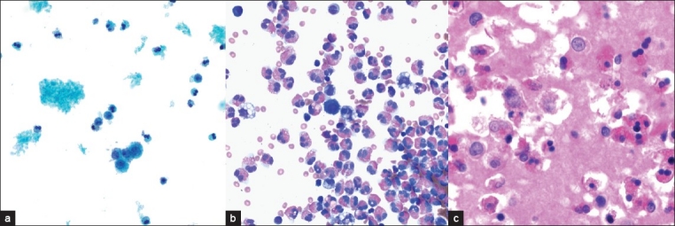
Background: There is a broad etiology for effusion eosinophilia that includes allergic, reactive, infectious, immune, neoplastic, and idiopathic causes. We report and describe the cytomorphologic findings of a rare case of eosinophilic ascites due to severe eosinophilic ileitis.
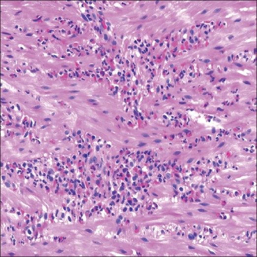
Case presentation: A 17-year-old male manifested acutely with eosinophilic ascites due to severe biopsy-proven subserosal eosinophilic ileitis. Isolated peritoneal fluid submitted for cytologic evaluation revealed that 65% eosinophils were present in a bloody background. The patient responded to corticosteroids, with complete resolution of his ascites.
Conclusion: Eosinophilic gastroenteritis with subserosal involvement should be added to the list of causes for eosinophils in peritoneal fluid. The finding of eosinophilic ascites, with appropriate clinical and laboratory findings, may warrant the need to perform laparoscopic intestinal biopsies to confirm the diagnosis.
Keywords: Ascites; cytology; eosinophilia; gastroenteritis; ileitis; peritoneal fluid; serosa.
Figures
Similar articles
-
Eosinophilic ascites as an uncommon presentation of eosinophilic gastroenteritis: A case report.Arab J Gastroenterol. 2021 Jun;22(2):184-186. doi: 10.1016/j.ajg.2021.02.002. Epub 2021 Jun 2. Arab J Gastroenterol. 2021. PMID: 34090834
-
Massive hemorrhagic ascites: A rare presentation of eosinophilic gastroenteritis.World J Clin Cases. 2018 Jul 16;6(7):156-160. doi: 10.12998/wjcc.v6.i7.156. World J Clin Cases. 2018. PMID: 30079343 Free PMC article.
-
Eosinophilic gastroenteritis as an uncommon cause of ascites recurrence in a young female.Med J Armed Forces India. 2023 Dec;79(Suppl 1):S283-S287. doi: 10.1016/j.mjafi.2021.10.005. Epub 2021 Nov 24. Med J Armed Forces India. 2023. PMID: 38144619 Free PMC article.
-
Causes of eosinophilic ascites - A systematic review.Rom J Intern Med. 2019 Jun 1;57(2):110-124. doi: 10.2478/rjim-2018-0041. Rom J Intern Med. 2019. PMID: 30864403
-
Eosinophilic gastroenteritis, ascites, and pancreatitis: a case report and review of the literature.South Med J. 2004 Sep;97(9):905-6. doi: 10.1097/01.SMJ.0000139403.55785.1C. South Med J. 2004. PMID: 15455985 Review.
Cited by
-
Authors attain comparable or slightly higher rates of citation publishing in an open access journal (CytoJournal) compared to traditional cytopathology journals - A five year (2007-2011) experience.Cytojournal. 2014 Apr 29;11:10. doi: 10.4103/1742-6413.131739. eCollection 2014. Cytojournal. 2014. PMID: 24987441 Free PMC article.
-
A review of uncommon cytopathologic diagnoses of pleural effusions from a chest diseases center in Turkey.Cytojournal. 2011;8:13. doi: 10.4103/1742-6413.83026. Epub 2011 Jul 16. Cytojournal. 2011. PMID: 21799700 Free PMC article.
-
Announcement of first time Cytojournal impact factor for 2012 coincides with Cytojournal decade celebration (2004-2013).Cytojournal. 2013 Aug 30;10:18. doi: 10.4103/1742-6413.117359. eCollection 2013. Cytojournal. 2013. PMID: 24082914 Free PMC article. No abstract available.
-
Intestinal Strongyloides causing peritoneal eosinophilia in peritoneal dialysis.Clin Kidney J. 2012 Dec;5(6):579-81. doi: 10.1093/ckj/sfs134. Epub 2012 Nov 6. Clin Kidney J. 2012. PMID: 26069806 Free PMC article.
References
-
- Bower G. Eosinophilic pleural effusion. A condition with multiple causes. Am Rev Respir Dis. 1967;95:746–51. - PubMed
-
- Galagan KA, Blomberg D, Cornbleet PJ, Glassy EF. Color Atlas of Body Fluids. An Illustrated. 3rd ed. Northfield, IL: College of American Pathologists; 2006. pp. 132–3.
-
- Kjeldsberg C, Knight J. Body fluids. 3rd ed. Chicago, IL: ASCP Press; 1993.
-
- Spriggs AI. Pleural eosinophilia due to pneumothorax. Acta Cytol. 1979;23:425. - PubMed
-
- De Smedt A, Vanderlinden E, Demanet C, De Waele M, Goossens A, Noppen M. Characterisation of pleural inflammation occurring after primary spontaneous pneumothorax. Eur Respir J. 2004;23:896–900. - PubMed
Publication types
LinkOut - more resources
Full Text Sources